
Quick Tips - Ultrasound Physics Doppler ShiftWhat is the arrow in the image referencing? You guessed it! Zero Doppler Shift - Let's talk about why. The Doppler effect or Doppler shift is the change in frequency of a wave in relation to an observer who is moving relative to the wave source. It is named after the Austrian physicist Christian Doppler, who described the phenomenon in 1842. In Color ultrasound the Doppler shift works with the ultrasound system to fill in color within the vessel when there are frequency changes in relation to the observer (transducer). When the direction of the sound beam is perpendicular to the direction of flow. There is no appreciable Doppler Shift and no color filling as a result. This is due to the cosine of the angle being 90 degrees.  Studying for your ultrasound registry exam? We can help! Our online ultrasound registry review course and Test & Learn Review Quiz can help you level up and pass your ultrasound registry exam today!
1 Comment
Quick Tips - GYN Ultrasound ANATOMY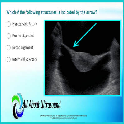 Which of the following structures is indicated by the arrow? Test your ultrasound anatomy skills!
You guessed it! This image is referring to the Broad Ligament. The broad ligament is a two-layered fold of peritoneum that extends from the sides of the uterus to the floor and lateral walls of the pelvis inferiorly and the adnexa superiorly. The broad ligament helps to hold the uterus in its anatomic position. It covers the uterus, ovaries, and fallopian tubes and also includes nerves and blood vessels to these organs. In this ultrasound image, the reason the broad ligament is easily identified is because of the fluid filling the pelvis. This highlights the location of the broad ligament. In normal settings, without a large amount of free fluid or the presence of other pathology, the broad ligament is rarely recognized on ultrasound. Studying for your OB/GYN ultrasound registry exam? Study with us! LEARN MORE Quick Ultrasound Physics Registry Review Tips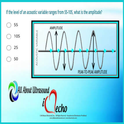 If the level of an acoustic variable ranges from 55-105, what is the amplitude? You guessed it! The answer is 25. But why? The amplitude is calculated by determining the median between the range values and then calculating the difference between the median and high/low values of the range. Amplitude is the amount of change in an acoustic variable. Amplitude is equal to the difference between average and the maximum or minimum values of an acoustic variable (or half the “peak-to-peak” amplitude). In the example in the question, the median of the range is 80. 80 is 25 above 55 and 25 below 105 - therefore the amplitude is equal to 25. The amplitude of ultrasound waves decrease as they travel through tissue, a phenomenon known as attenuation. For more info on amplitude and ultrasound physics, check out our other ultrasound physics resources. Studying for your ultrasound registry exam? We can help! Our ultrasound registry review courses and quizzes will take your ultrasound skills to the next level!
A Quick Look At Ovarian Doppler Waveforms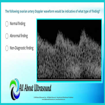 The following ovarian artery Doppler waveform would be indicative of what type of finding? The answer is ABNORMAL FINDING - but why? Let's take a quick look at the Doppler waveform and what makes it abnormal. The image reveals a low resistive waveform and is indicative of an abnormal ovarian Doppler finding. This can often be associated with Ovarian Torsion. When a blood vessel has a LOW RESISTIVE Doppler waveform appearance, this is due to the need for extra flow. Vascular beds that require a higher blood supply show these low resistive waveforms on spectral Doppler. This means that the waveform has a high diastolic component, indicating constant flow throughout diastole. This is common for vessels that supply muscles when you're working out, or the stomach and intestines when you've just eaten, or for the vessels that supply the brain and vital organs. However when a blood vessel shows LOW RESISTIVE characteristics for an organ that usually doesn't require a constant level of blood supply, this is a key marker that something isn't right and usually indicates stenosis. Blockage in the vessel will cause the vessel to become low resistive in order to compensate for the lack of flow.  Studying for your ultrasound registry exam? We can help! Our ultrasound registry review courses, quizzes will take your ultrasound skills to the next level!
|

Making Waves™All About Ultrasound presents Making Waves™, our ultrasound specific blog and newsletter. Join us here for ultrasound news, cases and more! Don't FORGET YOUR MERCH!
Archives
May 2023
Categories
All
|
 RSS Feed