ULTRASOUND REGISTRY REVIEW SIMPLIFIED
Picture
   

Making Waves™ - All About Ultrasound BloG

  • Home
  • Ultrasound Services
    • Ultrasound Accreditation
    • Competency Assessments
  • Ultrasound Education
    • E-Learning >
      • E-Learning & CME
      • Upper Extremity Duplex
      • Venous Insufficiency
      • Doppler Principles and Hemodynamics
      • Left Ventricular Diastology
      • Constrictive Pericarditis
      • Aortic Stenosis
      • Ultrasound 101
      • Echo Basics 101
    • Free Membership
    • On-Site Training
    • Scanning Quick Guides
  • Ultrasound Registry Review
    • Free Membership
    • Upcoming Events
    • Ultrasound Physics SPI
    • Advanced Cardiac Sonographer
    • Adult Echocardiography
    • Pediatric Echocardiography
    • Fetal Echocardiography
    • Vascular Ultrasound
    • Abdomen Ultrasound
    • OB-GYN Ultrasound
    • Breast Ultrasound
    • Group | Team Access
  • Ultrasound Jobs
  • Ultrasound Resources
    • Ultrasound Case Studies
    • Sonography Coloring Book
    • Ultrasound Worksheets
    • Ultrasound Protocols
    • Ultrasound Credentialing
    • ULTRASOUND SCHOOLS
    • Sonography News
  • Making Waves™ Blog
  • SONO SHOP
  • Newsletter
  • FAQ
  • About Us
  • Contact Us
  • FREE SIGN UP
  • LOGIN
  • iHeartEcho

11/4/2022

Quick Physics Tips Amplitude

0 Comments

Read Now
 

Quick Ultrasound Physics Registry Review Tips
Picture
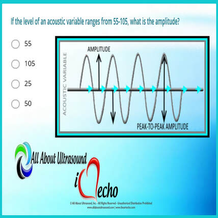
If the level of an acoustic variable ranges from 55-105, what is the amplitude?

You guessed it! The answer is 25. But why?

The amplitude is calculated by determining the median between the range values and then calculating the difference between the median and high/low values of the range.

Amplitude is the amount of change in an acoustic variable. Amplitude is equal to the difference between average and the maximum or minimum values of an acoustic variable (or half the “peak-to-peak” amplitude).

In the example in the question, the median of the range is 80. 
​80 is 25 above 55 and 25 below 105 - therefore the amplitude is equal to 25. 

The amplitude of ultrasound waves decrease as they travel through tissue, a phenomenon known as attenuation. For more info on amplitude and ultrasound physics, check out our other ultrasound physics resources. 

Picture
Studying for your ultrasound registry exam? We can help! Our ultrasound registry review courses and quizzes will take your ultrasound skills to the next level! ​
Learn More

Share

0 Comments


Leave a Reply.

Details
    Picture

    Making Waves​™

    All About Ultrasound presents Making Waves™, our ultrasound specific blog and newsletter. Join us here for ultrasound news, cases and more! 

    Suggestions or Comments?
    Tell us your opinion...
    [email protected]

    Don't FORGET YOUR MERCH!

    SHOP NOW
    Picture

    Archives

    May 2023
    April 2023
    November 2022
    July 2020
    June 2020
    January 2019
    December 2018
    February 2018
    January 2018
    July 2017
    June 2017
    May 2017
    April 2017
    February 2017
    January 2017
    September 2016
    August 2016
    July 2016
    June 2016
    May 2016
    May 2015
    April 2015
    March 2015
    January 2015
    July 2014
    July 2013
    March 2013
    February 2013
    November 2012
    April 2012
    April 2011
    March 2011
    January 2011
    November 2010

    Categories

    All
    Accreditation
    Case Studies
    Echocardiography
    General
    Job Search
    OB/GYN
    Physics
    Registries
    Salary
    Scanning Tips
    Ultrasound News
    Ultrasound Research
    Vascular
    Webinar

    RSS Feed


ALL RIGHTS RESERVED
​​©ALL ABOUT ULTRASOUND, INC. dba iHEARTECHO

TERMS OF SERVICE   ●   PRIVACY POLICY   ●   
SUPPORT
​
[email protected]

ARDMS® is a registered trademark of the American Registry for Diagnostic Medical Sonography and is not associated with this website.  
CCI® is a registered trademark of Cardiovascular Credentialing International and is not associated with this website.
​
​WEB DESIGN BY: SoundMedia
  • Home
  • Ultrasound Services
    • Ultrasound Accreditation
    • Competency Assessments
  • Ultrasound Education
    • E-Learning >
      • E-Learning & CME
      • Upper Extremity Duplex
      • Venous Insufficiency
      • Doppler Principles and Hemodynamics
      • Left Ventricular Diastology
      • Constrictive Pericarditis
      • Aortic Stenosis
      • Ultrasound 101
      • Echo Basics 101
    • Free Membership
    • On-Site Training
    • Scanning Quick Guides
  • Ultrasound Registry Review
    • Free Membership
    • Upcoming Events
    • Ultrasound Physics SPI
    • Advanced Cardiac Sonographer
    • Adult Echocardiography
    • Pediatric Echocardiography
    • Fetal Echocardiography
    • Vascular Ultrasound
    • Abdomen Ultrasound
    • OB-GYN Ultrasound
    • Breast Ultrasound
    • Group | Team Access
  • Ultrasound Jobs
  • Ultrasound Resources
    • Ultrasound Case Studies
    • Sonography Coloring Book
    • Ultrasound Worksheets
    • Ultrasound Protocols
    • Ultrasound Credentialing
    • ULTRASOUND SCHOOLS
    • Sonography News
  • Making Waves™ Blog
  • SONO SHOP
  • Newsletter
  • FAQ
  • About Us
  • Contact Us
  • FREE SIGN UP
  • LOGIN
  • iHeartEcho