ULTRASOUND REGISTRY REVIEW SIMPLIFIED
Picture
   

Making Waves™ - All About Ultrasound BloG

  • Home
  • Ultrasound Services
    • Ultrasound Accreditation
    • Competency Assessments
  • Ultrasound Education
    • E-Learning >
      • E-Learning & CME
      • Upper Extremity Duplex
      • Venous Insufficiency
      • Doppler Principles and Hemodynamics
      • Left Ventricular Diastology
      • Constrictive Pericarditis
      • Aortic Stenosis
    • Free Membership
    • On-Site Training
    • Scanning Quick Guides
  • Ultrasound Registry Review
    • Free Membership
    • Upcoming Events
    • Ultrasound Physics SPI
    • Advanced Cardiac Sonographer
    • Adult Echocardiography
    • Pediatric Echocardiography
    • Fetal Echocardiography
    • Vascular Ultrasound
    • Abdomen Ultrasound
    • OB-GYN Ultrasound
    • Breast Ultrasound
    • Group | Team Access
  • Ultrasound Jobs
  • Ultrasound Resources
    • Ultrasound Case Studies
    • Sonography Coloring Book
    • Ultrasound Worksheets
    • Ultrasound Protocols
    • Ultrasound Credentialing
    • ULTRASOUND SCHOOLS
    • Sonography News
  • Making Waves™ Blog
  • SONO SHOP
  • Newsletter
  • FAQ
  • About Us
  • Contact Us
  • FREE SIGN UP
  • LOGIN
  • iHeartEcho

7/20/2017

5 Best Practice Tips to Ob-Gyn Ultrasound

16 Comments

Read Now
 
1. That Bladder! We have all seen the pregnant pee pee dance… While sometimes uncomfortable for our patients, this is the key to the first trimester transabdominal approach and also to 2nd trimester cervical measurements. Sometimes it just can’t be avoided, your patient is going to dance! However, in the 2nd trimester, it can be helpful for your patient to get what you need with the bladder full and to let them empty it out before completing the rest of your study. Often this can also improve the position of the fetus and help you to get better images with a relaxed mama and a relaxed baby!
Picture
Picture
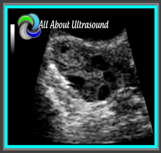
2. Scanning and trying to find those ovaries can prove to be a challenge with a pregnant uterus! Even though it is not our focus, we can’t forget those ovaries! Making sure to image those along with the uterus and fetal images will ensure that you’re not missing potential pathology, but it can definitely be tough with that pregnant uterus in the way. Best methods are to bring the transducer laterally to the patient’s side and to scan in a transverse plane along the lateral side of the uterus, starting pretty high up and moving down toward the patient’s cervix. That transverse plane will give you an increased field of view and will allow you to identify structures more easily.

3. Outflow tracts… the sound of that might scare some sonographers who are not used to looking at the fetal heart or evaluating this on a regular basis, but it’s much easier than you might think. Remember there are two main blood vessels that exit the heart – the aorta and the pulmonary artery. That’s it… just two. So how do you tell which is which? Most of the time (unless in a case of transposition of the great arteries or double outlet right ventricle), the aorta exits the left ventricle and the pulmonary artery exits the right ventricle. It’s important to note that the aorta will arch around after it leaves the heart and extend into the thoracic/abdominal aorta. The pulmonary artery will typically split into two branches.

​Yes, there are many variations that can happen with cardiac pathology and unless you specialize in maternal fetal medicine or fetal echocardiography, you may not know the specifics of every congenital heart disease you might encounter, but if you know the basics and what that looks like, you should be able to know when it’s just not right and alert your reading physician. If you’re not scanning this on a regular basis, it would be a good idea to add this to your daily protocol.
Picture
Picture
Picture
4. Amniotic Fluid Index – how do you know your measurements are accurate? Many sonographers are not aware of how to properly measure fluid for an AFI. One key tip is to scan with color Doppler flow on – this allows you to identify umbilical cord that might be floating within the pocket of fluid. Another best practice tip and proper way to perform AFI measurements is to be sure your patient is lying flat on the table and keep the probe/ultrasound beam perpendicular to the floor. Also, keep your measurements perpendicular as well. This helps to ensure that the fluid pocket is not falsely enlarged.
5. Gender! We’ve all been there, where it seems that everyone is there for the show. While somehow you ended up being the entertainment for the expectant parents, don’t forget how exciting it can be to know how to plan for your little one. Yes, it’s often a pain with the dad, grandma, grandpa, sister, brother, niece, nephew, the mailman, the neighbor, the sister’s boyfriend’s mother-in-law and everyone else trying to squeeze into the room to catch a glance of the precious cargo and whatever package the baby might be sporting… so take a deep breath, don’t lose it on grandma and remember to SMILE 😊. Oh and scanning, well you know how that goes…​ get those pesky diagnostic measurements and anatomy evaluations out of the way and  then... turtle or hamburger… take your pick!
Picture
Picture

​

Share

16 Comments

4/10/2012

Parents Focusing on Gender

4 Comments

Read Now
 
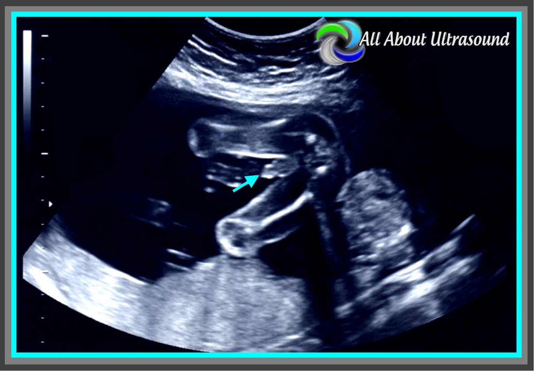
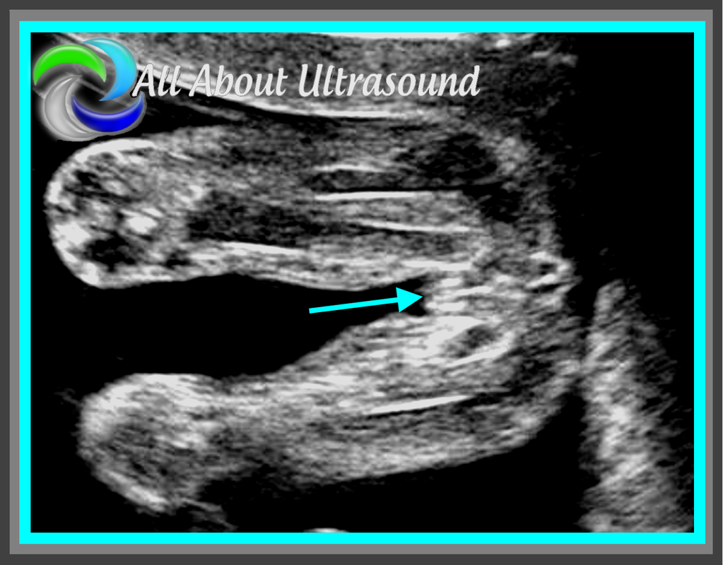
For our OB/GYN Sonographers: How do you handle it when parents-to-be are so focused on the gender of their baby that they forget they are having their ultrasound to determine the health of their baby?  So many times parents can be so excited and only want to know the sex of their baby. This can make it difficult to focus on the anatomical survey you as the sonographer are there to perform. How do you handle this professionally? Also, for those times when there is actually something wrong with the baby, then what? How do you maintain your professionalism as a sonographer, but still make it clear that your focus is not the baby's gender?

Share

4 Comments
Details
    Picture

    Making Waves​™

    All About Ultrasound presents Making Waves™, our ultrasound specific blog and newsletter. Join us here for ultrasound news, cases and more! 

    Suggestions or Comments?
    Tell us your opinion...
    [email protected]

    Don't FORGET YOUR MERCH!

    SHOP NOW
    Picture

    Archives

    May 2023
    April 2023
    November 2022
    July 2020
    June 2020
    January 2019
    December 2018
    February 2018
    January 2018
    July 2017
    June 2017
    May 2017
    April 2017
    February 2017
    January 2017
    September 2016
    August 2016
    July 2016
    June 2016
    May 2016
    May 2015
    April 2015
    March 2015
    January 2015
    July 2014
    July 2013
    March 2013
    February 2013
    November 2012
    April 2012
    April 2011
    March 2011
    January 2011
    November 2010

    Categories

    All
    Accreditation
    Case Studies
    Echocardiography
    General
    Job Search
    OB/GYN
    Physics
    Registries
    Salary
    Scanning Tips
    Ultrasound News
    Ultrasound Research
    Vascular
    Webinar

    RSS Feed


ALL RIGHTS RESERVED
​​©ALL ABOUT ULTRASOUND, INC. dba iHEARTECHO

TERMS OF SERVICE   ●   PRIVACY POLICY   ●   
SUPPORT
​
[email protected]

ARDMS® is a registered trademark of the American Registry for Diagnostic Medical Sonography and is not associated with this website.  
CCI® is a registered trademark of Cardiovascular Credentialing International and is not associated with this website.
​
​WEB DESIGN BY: SoundMedia
  • Home
  • Ultrasound Services
    • Ultrasound Accreditation
    • Competency Assessments
  • Ultrasound Education
    • E-Learning >
      • E-Learning & CME
      • Upper Extremity Duplex
      • Venous Insufficiency
      • Doppler Principles and Hemodynamics
      • Left Ventricular Diastology
      • Constrictive Pericarditis
      • Aortic Stenosis
    • Free Membership
    • On-Site Training
    • Scanning Quick Guides
  • Ultrasound Registry Review
    • Free Membership
    • Upcoming Events
    • Ultrasound Physics SPI
    • Advanced Cardiac Sonographer
    • Adult Echocardiography
    • Pediatric Echocardiography
    • Fetal Echocardiography
    • Vascular Ultrasound
    • Abdomen Ultrasound
    • OB-GYN Ultrasound
    • Breast Ultrasound
    • Group | Team Access
  • Ultrasound Jobs
  • Ultrasound Resources
    • Ultrasound Case Studies
    • Sonography Coloring Book
    • Ultrasound Worksheets
    • Ultrasound Protocols
    • Ultrasound Credentialing
    • ULTRASOUND SCHOOLS
    • Sonography News
  • Making Waves™ Blog
  • SONO SHOP
  • Newsletter
  • FAQ
  • About Us
  • Contact Us
  • FREE SIGN UP
  • LOGIN
  • iHeartEcho