ULTRASOUND REGISTRY REVIEW SIMPLIFIED
Picture
   

Making Waves™ - All About Ultrasound BloG

  • Home
  • Ultrasound Services
    • Ultrasound Accreditation
    • Competency Assessments
  • Ultrasound Education
    • E-Learning >
      • E-Learning & CME
      • Upper Extremity Duplex
      • Venous Insufficiency
      • Doppler Principles and Hemodynamics
      • Left Ventricular Diastology
      • Constrictive Pericarditis
      • Aortic Stenosis
    • Free Membership
    • On-Site Training
    • Scanning Quick Guides
  • Ultrasound Registry Review
    • Free Membership
    • Upcoming Events
    • Ultrasound Physics SPI
    • Advanced Cardiac Sonographer
    • Adult Echocardiography
    • Pediatric Echocardiography
    • Fetal Echocardiography
    • Vascular Ultrasound
    • Abdomen Ultrasound
    • OB-GYN Ultrasound
    • Breast Ultrasound
    • Group | Team Access
  • Ultrasound Jobs
  • Ultrasound Resources
    • Ultrasound Case Studies
    • Sonography Coloring Book
    • Ultrasound Worksheets
    • Ultrasound Protocols
    • Ultrasound Credentialing
    • ULTRASOUND SCHOOLS
    • Sonography News
  • Making Waves™ Blog
  • SONO SHOP
  • Newsletter
  • FAQ
  • About Us
  • Contact Us
  • FREE SIGN UP
  • LOGIN
  • iHeartEcho

7/16/2020

5 Quick Scanning Tips For Carotid Ultrasound

1 Comment

Read Now
 
Scanning those tortuous Carotid vessels can sometimes be tricky! So here we'll go through 5 quick scanning tips to help you get great carotid images and accurate velocities. 
Picture
1. Do a Quick Scan Survey
Taking a quick look through the entire vessel and bifurcation before you start imaging will help you know of any plaque that might be present and will also help you be aware of the vessel course. This will help you better prepare to angle your Color & Doppler through any stenosis and also if there are tortuous vessels. It is always a good idea to know what you are getting yourself into first. 
2. Be Consistent
Often times your scanning protocol will be determined by the facility you work for. This is helpful with accreditation and standardization to make sure that accurate and consistent exams are performed every time, for every patient. With regard to protocol, it can sometimes be difficult to jump around from one vessel location, like proximal to the distal and then ECA and ICA and then go back to obtain it again for other imaging modes like Color and Spectral Doppler. So if at all possible, it is best to stay consistent, perform all imaging modes (2D, Color and PW Doppler) in that one location and then move on. 
Picture
Picture
3. Heel/Toe Will Come in Handy
Using the "Heel/Toe" method to rock and tilt the angle of the transducer will help to line up the vessel to the correct Doppler angle. This is especially useful when there is "deep dive" of the vessel or when there is a tortuous curve. By using your hand to angle the transducer, you can maintain that 60 degree angle and ensure that each Doppler sample is angled correctly. 
4. It's All About the Angle
Remember your Doppler angle should be parallel to blood flow through the vessel (not the vessel wall). This is important when there is a stenosis, because often the flow direction will change due to the shape of the plaque. Also, it's important to remember that the SVU recommends keeping your Doppler angle between 45-60 degrees in order to maintain consistency in reporting velocities. It can be helpful if you are doing a follow up study for a patient with a known stenosis to check the prior ultrasound images (not just the report) and review the Doppler angle used previously and where the stenosis was sampled. This can help to reduce errors between sonographers and ensure that the velocities reported are consistent and only different if there is truly a reduction in vessel lumen since the previous exam. 
Picture
Picture
5. Attention to Detail
Sometimes those tortuous vessels and deep vessels can be difficult. Pay attention to your PRF/scale, your wall filter settings and your color box angle. This can help bring in flow in those difficult exams. Often steering your color box straight will help to pick up those deep diving vessels. 

Get Our FREE SCANNING TIP DOWNLOAD
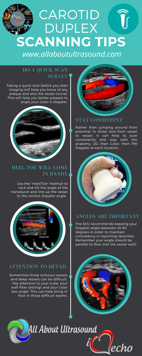
FREE DOWNLOAD
Picture

Share

1 Comment

7/29/2017

Step by Step Guide to Mesenteric Abdominal Duplex

6 Comments

Read Now
 
Picture
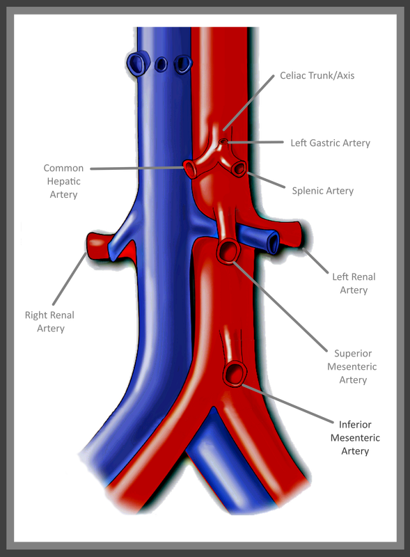
Mesenteric Abdominal Duplex – it sounds complicated right? In reality though, as long as your patient is prepped, there’s not much to it. I say that with caution though – we’ve all had those patients that just really need another modality of testing. Let's be real... we all know that our ultrasound transducer is NOT a magic wand! 

First and foremost - Patient Prep! Your patient prep is one of the most important factors when performing Mesenteric Duplex Ultrasound. Your patient needs to be NPO for at least 6-8 hours before scanning. A test done without the right prep, might as well have not been done at all.  This is for a number of reasons:
1. You can't see squat with air and gas in the way!
2. The mesenteric arterial system should be scanned both pre and post prandial for evaluation of stenosis and arterial ischemic response. ​

Picture
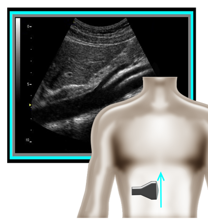
​So where to begin and what protocol? I've got that covered for you! A mesenteric duplex protocol should include, at a minimum...  Transverse and Longitudinal approach with 2D, Longitudinal approach with Color and PW Doppler (Record PSV and EDV) at each of the following locations:                                                                                                                                                       
•Proximal Aorta
•Celiac Artery/Trunk
•Branching of Common Hepatic Artery and Splenic Artery from the Celiac Artery
•SMA origin, proximal, mid, distal
•IMA origin, proximal and as distal as possible
•IVC 

​A few things can help you improve your imaging and will also help you evaluate patient pathology.
Picture
Picture
1. Patient position is an important factor to remember. You should position your patient in a slightly elevated/supine position. This allows for a better view with less pressure on the diaphragm. Another trick is to have your patient lay supine and bend their knees. This also reduces pressure and tension on the diaphragm and allows for better visualization of the structures near the xyphoid process (ie. proximal aorta, celiac trunk, SMA). Having the patient sit erect can also help with evaluation of median arcuate ligament compression and if images are limited otherwise.
​
​2. Using patient respirations to control the movement of structures and improve image quality (especially for Doppler waveforms). Respirations also help to evaluate for median arcuate ligament compression is sometimes a major factor in diagnosing this condition. Patient's with median arcuate ligament compression will have mildly elevated flow in the Celiac Artery with inspiration (this is normal). However, with expiration, the artery will be compressed by the median arcuate ligament and will cause an extrinsic obstruction due to compression and a marked increase in flow velocity. Using patient respirations to assess for this is a major factor of the Mesenteric Duplex exam. 

​3. Use Power Doppler - on those difficult patients, power Doppler can be your savior! This can also be used to visualize the Celiac Branches, the IMA and other smaller vessels.
Picture
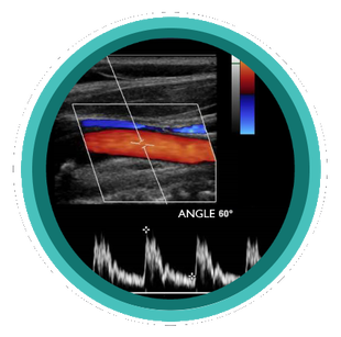
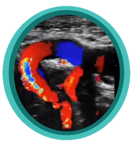
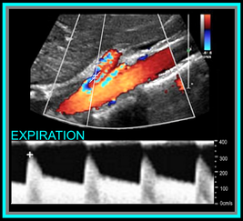
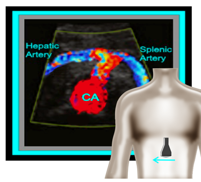
Okay, so now that we have the basics out of the way... you're probably wondering, what should the waveforms look like and what is normal/abnormal? So here's a quick guide:

​Celiac Artery
​

Remember that the Celiac Artery supplies the liver, spleen and stomach, which are low resistance vascular beds. Normal Doppler waveforms will show increased diastolic flow because of the organs supplied. Flow may also increase with inspiration. ​

Don't forget to evaluate the branches! This is best done in a transverse plane. 

≥70% Celiac Artery Stenosis will show a peak systolic velocity of  ≥200 cm/s.
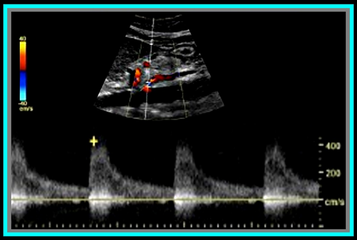
Picture
​Superior Mesenteric Artery
​

​Remember that the SMA supplies the jejunum, ileum, and both the right and transverse colon. Because of this, waveform characteristics will vary based on state (ie. NPO, post-prandial). Diastolic flow will increase as needed for digestion, post-prandially.

In the normal vessel, post-prandial evaluation should show increased peak systolic flow velocities. If PSV flow does not increase, this is suggestive of a hemodynamically significant stenosis. Also keep in mind that inspiration will show an increase in peak systolic velocities.

≥70% SMA stenosis will show a peak systolic velocity of  ≥275cm/s or absence of color flow in the SMA. End diastolic flow velocities of ≥45cm/s are also an indication of ≥70% SMA stenosis. 

​Also keep in mind that you can find pathology based on the angle of the SMA  takeoff from the aorta. If the angle is markedly increased, it may indicate the presence of adenopathy. The SMA should course parallel to the aorta. 


Inferior Mesenteric Artery

​The IMA supplies the distal 1/3 of the transverse colon, splenic flexure, descending colon, sigmoid colon and rectum. It's waveform characteristics are similar to the SMA and will vary based on state (ie. NPO, post-prandial). Diastolic flow will increase as needed for digestion, post-prandially.

≥70% IMA stenosis will show a peak systolic velocity of  ≥275cm/s or absence of color flow in the IMA.

​You may need to get your magic wand out for this one! 
Picture
Diagnostic Criteria Reference: Moneta, et al

GET OUR FREE SCANNING TIPS QUICK GUIDE

FREE DOWNLOAD
Picture

Share

6 Comments

3/23/2011

Ultrasound Accreditation

3 Comments

Read Now
 
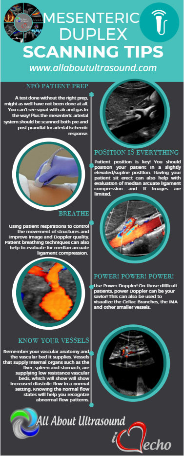
Ultrasound Accreditation and/or sonographer credentialing requirements are already in effect in many states and soon will be required for reimbursement of exams. Is your ultrasound lab accredited? If not, are the sonographers in your department registered? How is your lab preparing for the upcoming accreditation requirements?

Share

3 Comments

11/21/2010

Internal Carotid Occlusion

3 Comments

Read Now
 
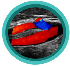
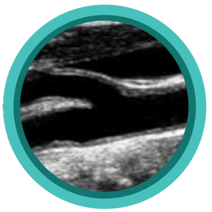
How would you adjust your ultrasound settings to go about proving this diagnosis? What waveform characteristics would you be aware of in adjacent vasculature?
Picture
Picture
Picture
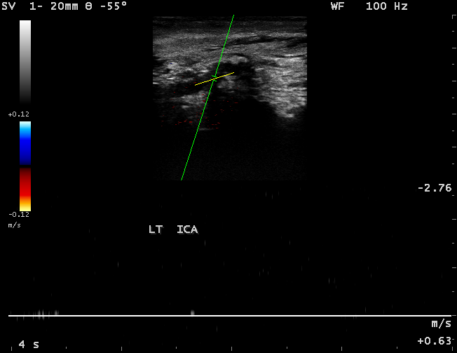
Images courtesy of SONA IMAGING SOLUTIONS

Share

3 Comments
Details
    Picture

    Making Waves​™

    All About Ultrasound presents Making Waves™, our ultrasound specific blog and newsletter. Join us here for ultrasound news, cases and more! 

    Suggestions or Comments?
    Tell us your opinion...
    [email protected]

    Don't FORGET YOUR MERCH!

    SHOP NOW
    Picture

    Archives

    May 2023
    April 2023
    November 2022
    July 2020
    June 2020
    January 2019
    December 2018
    February 2018
    January 2018
    July 2017
    June 2017
    May 2017
    April 2017
    February 2017
    January 2017
    September 2016
    August 2016
    July 2016
    June 2016
    May 2016
    May 2015
    April 2015
    March 2015
    January 2015
    July 2014
    July 2013
    March 2013
    February 2013
    November 2012
    April 2012
    April 2011
    March 2011
    January 2011
    November 2010

    Categories

    All
    Accreditation
    Case Studies
    Echocardiography
    General
    Job Search
    OB/GYN
    Physics
    Registries
    Salary
    Scanning Tips
    Ultrasound News
    Ultrasound Research
    Vascular
    Webinar

    RSS Feed


ALL RIGHTS RESERVED
​​©ALL ABOUT ULTRASOUND, INC. dba iHEARTECHO

TERMS OF SERVICE   ●   PRIVACY POLICY   ●   
SUPPORT
​
[email protected]

ARDMS® is a registered trademark of the American Registry for Diagnostic Medical Sonography and is not associated with this website.  
CCI® is a registered trademark of Cardiovascular Credentialing International and is not associated with this website.
​
​WEB DESIGN BY: SoundMedia
  • Home
  • Ultrasound Services
    • Ultrasound Accreditation
    • Competency Assessments
  • Ultrasound Education
    • E-Learning >
      • E-Learning & CME
      • Upper Extremity Duplex
      • Venous Insufficiency
      • Doppler Principles and Hemodynamics
      • Left Ventricular Diastology
      • Constrictive Pericarditis
      • Aortic Stenosis
    • Free Membership
    • On-Site Training
    • Scanning Quick Guides
  • Ultrasound Registry Review
    • Free Membership
    • Upcoming Events
    • Ultrasound Physics SPI
    • Advanced Cardiac Sonographer
    • Adult Echocardiography
    • Pediatric Echocardiography
    • Fetal Echocardiography
    • Vascular Ultrasound
    • Abdomen Ultrasound
    • OB-GYN Ultrasound
    • Breast Ultrasound
    • Group | Team Access
  • Ultrasound Jobs
  • Ultrasound Resources
    • Ultrasound Case Studies
    • Sonography Coloring Book
    • Ultrasound Worksheets
    • Ultrasound Protocols
    • Ultrasound Credentialing
    • ULTRASOUND SCHOOLS
    • Sonography News
  • Making Waves™ Blog
  • SONO SHOP
  • Newsletter
  • FAQ
  • About Us
  • Contact Us
  • FREE SIGN UP
  • LOGIN
  • iHeartEcho