ULTRASOUND REGISTRY REVIEW SIMPLIFIED
Picture
   

Making Waves™ - All About Ultrasound BloG

  • Home
  • Ultrasound Services
    • Ultrasound Accreditation
    • Competency Assessments
  • Ultrasound Education
    • E-Learning >
      • E-Learning & CME
      • Upper Extremity Duplex
      • Venous Insufficiency
      • Doppler Principles and Hemodynamics
      • Left Ventricular Diastology
      • Constrictive Pericarditis
      • Aortic Stenosis
    • Free Membership
    • On-Site Training
    • Scanning Quick Guides
  • Ultrasound Registry Review
    • Free Membership
    • Upcoming Events
    • Ultrasound Physics SPI
    • Advanced Cardiac Sonographer
    • Adult Echocardiography
    • Pediatric Echocardiography
    • Fetal Echocardiography
    • Vascular Ultrasound
    • Abdomen Ultrasound
    • OB-GYN Ultrasound
    • Breast Ultrasound
    • Group | Team Access
  • Ultrasound Jobs
  • Ultrasound Resources
    • Ultrasound Case Studies
    • Sonography Coloring Book
    • Ultrasound Worksheets
    • Ultrasound Protocols
    • Ultrasound Credentialing
    • ULTRASOUND SCHOOLS
    • Sonography News
  • Making Waves™ Blog
  • SONO SHOP
  • Newsletter
  • FAQ
  • About Us
  • Contact Us
  • FREE SIGN UP
  • LOGIN
  • iHeartEcho

6/11/2020

Constrictive Pericarditis

2 Comments

Read Now
 
Picture

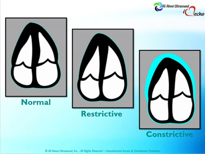
​​​Mastering Echocardiography can be tough, especially when it comes to complex processes like Constrictive Pericarditis. Patients with Constrictive Pericarditis do not present for typical pericarditis symptoms. Instead, they present with symptoms of heart failure and so this can often be a challenge for even very skilled sonographers to identify the subtle differences between Restrictive Cardiomyopathy and Constrictive Pericarditis. These two disease processes can appear very similar on echocardiography. However, there are a few things that clue us into the correct diagnosis.

Picture
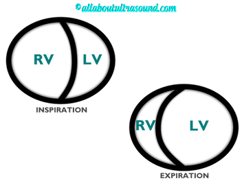
Constrictive Pericarditis is seen with a fibrotic, thickened pericardium, which insulates the ventricle and constricts the ventricle from fully relaxing during diastole. Whereas, Restrictive Cardiomyopathy results in a thickened myocardium, which inhibits the ability of the ventricle to relax and also results in abnormal diastolic function. However, that's where the similarities end. ​​
 
​​First of all, we need to evaluate for interdependence of the ventricles. But what does that mean? Ventricular interdependence is when there is a respiratory ventricular septal shift. This then leads to an increase in the volume of one ventricle associated with a decreased volume in the opposite ventricle. This can be tricky to diagnose, so it takes a pretty detailed echo exam and a keen eye of the sonographer and physician. 

Picture
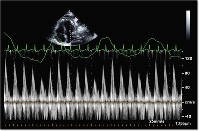
Secondly, when evaluating for constriction, we will need to look at respiratory changes to the Doppler waveform patterns. The respiratory changes in the Mitral inflow pattern will show a variation of greater than 15% when constriction is present. Whereas, with restriction, this waveform pattern will show a restrictive filling pattern with an E/A ratio >2.0 and deceleration time <160ms.


Picture
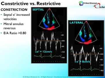
Another key factor in determining constriction vs. restriction is the E/e' ratio and diastolic function. Remember that both disease processes will have a degree of diastolic dysfunction. ​Normal left ventricular function will typically show a lateral e’ greater than septal/medial e' velocities, because the septal wall is somewhat restricted and the lateral wall is more free to move. However, with constriction the pericardium is insulating the ventricular movements and does not allow for full relaxation and movement of the lateral wall. This results in mitral annulus reversus, which is a decreased lateral e' velocity and compensatory increase in tissue velocities in the septal/medial annulus. Restrictive patterns in tissue velocities will show an overall decrease in both the lateral and septal e' velocities.

Picture
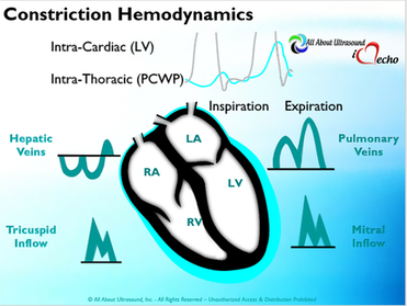
Additionally, with constriction, this will result in expiratory hepatic vein reversal. But why does this occur? Remember that air moves from areas of high pressure to low pressure, which allows for air flow into the lungs. During expiration, the volume of air (and the pressure) of the thoracic cavity decreases, causing the intrapulmonary pressure to rise above the atmospheric pressure. However, with constriction, the pericardium is insulating the intracardiac chambers and this keeps them from tracking normally with intrapulmonary pressures. Remember the pulmonary artery and aorta are OUTSIDE of the pericardium. 

​So with constriction the thorax pressure and the  pulmonary venous pressure will drop, but there are phasic filling differences within the heart, because the gradient to fill the left side of the heart is decreased. The heart is no longer able to push outward against the pericardium, so the pressure is forced inward into the cardiac chambers. So during expiration, the pressure in the right atrium causes flow reversal in the hepatic veins. Whereas, with chronic late stages of restriction, there is an inspiratory reversal during diastole, because there is no shifting of septum and the right heart cannot accommodate increased flow which results from chronic diastolic dysfunction and this causes hepatic vein reversal. 

W​hile diagnosing Constrictive Pericarditis on echocardiography can certainly be a challenge, it can be done with confidence. Paying close attention to the details and recognizing when your patient with heart failure symptoms might actually be more than meets the eye, can help to make the correct diagnosis and ensure adequate treatment for your patient. 
Want more information on the differences between Constrictive Pericarditis and Restrictive Cardiomyopathy, including Strain Imaging methods? See our E-Learning Course Mastering Constrictive Pericarditis and earn 1 SDMS CME credit. ​
Enroll Now For CME
Re-post from: ​https://www.iheartecho.com/echoblog/constrictive-pericarditis

REFERENCES
  • Ling LH, Oh JK, Schaff HV et al. Constrictive pericarditis in the modern era: evolving clinical spectrum and impact on outcome after pericardiectomy. Circulation. 1999;100(13): 1380-6.
  • Myers RB, Spodick DH. Constrictive pericarditis: clinical and pathophysiologic characteristics. Am Heart J. 1999;138(2 Pt 1):219-32.
  • Mehta A, Mehta M, Jain AC. Constrictive pericarditis. Clin Cardiol. 1999;22(5):334-44.
  • Hancock EW. Differential diagnosis of restrictive cardiomyopathy and constrictive pericarditis. Heart. 2001;86(3): 343-9.
  • Ling LH, Oh JK, Tei C, et al. Pericardial thickness measured with transesophageal echocardiography: feasibility and potential clinical usefulness. J Am Coll Cardiol. 1997; 29(6):1317-23.
  • Talreja DR, Edwards WD, Danielson GK, et al. Constrictive pericarditis in 26 patients with histologically normal pericardial thickness. Circulation. 2003;108(15):1852-7.
  • Rajagopalan N, Garcia MJ, Rodriguez L, et al. Comparison of new Doppler echocardiographic methods to differentiate constrictive pericardial heart disease and restrictive cardiomyopathy. Am J Cardiol. 2001;87(1):86-94.
  • Oh JK, Hatle LK, Seward JB, et al. Diagnostic role of Doppler echocardiography in constrictive pericarditis. J Am Coll Cardiol. 1994;23(1):154-62.
  • Hatle LK, Appleton CP, Popp RL. Differentiation of constrictive pericarditis and restrictive cardiomyopathy by Doppler echocardiography. Circulation. 1989;79(2):357-70.
  • Oh JK, Tajik AJ, Appleton CP, et al. Preload reduction to unmask the characteristic Doppler features of constrictive pericarditis. A new observation. Circulation. 1997; 95(4):796-9.
  • Sengupta PP, Mohan JC, Mehta V et al. Accuracy and pitfalls of early diastolic motion of the mitral annulus for diagnosing constrictive pericarditis by tissue Doppler imaging. Am J Cardiol. 2004;93(7):886-90.
  • Ha JW, Ommen SR, Tajik AJ, et al. Differentiation of constrictive pericarditis from restrictive cardiomyopathy using mitral annular velocity by tissue Doppler echocardiography. Am J Cardiol. 2004;94(3):316-9.
  • Garcia MJ, Rodriguez L, Ares M, et al. Differentiation of constrictive pericarditis from restrictive cardiomyopathy: assessment of left ventricular diastolic velocities in longitudinal axis by Doppler tissue imaging. J Am Coll Cardiol. 1996;27(1):108-14.
  • von Bibra H, Schober K, Jenni R, et al. Diagnosis of constrictive pericarditis by pulsed Doppler echocardiography of the hepatic vein. Am J Cardiol. 1989;63(7):483-8.
  • Ha JW, Oh JK, Ling LH, et al. Annulus paradoxus: transmitral flow velocity to mitral annular velocity ratio is inversely proportional to pulmonary capillary wedge pressure in patients with constrictive pericarditis. Circulation. 2001;104(9):976-8.
  • Reuss CS, Wilansky SM, Lester SJ, et al. Using mitral 'annulus reversus' to diagnose constrictive pericarditis. Eur J Echocardiogr. 2009;10(3):372-5.
  • Sengupta PP, Mohan JC, Mehta V et al. Doppler tissue imaging improves assessment of abnormal interventricular septal and posterior wall motion in constrictive pericarditis. J Am Soc Echocardiogr. 2005;18(3):226-30.
  • Sengupta PP, Krishnamoorthy VK, Abhayaratna WP, et al. Disparate patterns of left ventricular mechanics differentiate constrictive pericarditis from restrictive cardiomyopathy. JACC Cardiovasc Imaging. 2008;1(1):29-38.
  • Circ Cardiovasc Imaging. 2014 May;7(3):526-34. doi: 10.1161/CIRCIMAGING.113.001613. Epub 2014 Mar 14.
  • American Society of Echocardiography Clinical Recommendations for Multimodality Cardiovascular Imaging of Patients with Pericardial Disease (J Am Soc Echocardiogr 2013;26:965-1012.)


Share

2 Comments
Details
    Picture

    Making Waves​™

    All About Ultrasound presents Making Waves™, our ultrasound specific blog and newsletter. Join us here for ultrasound news, cases and more! 

    Suggestions or Comments?
    Tell us your opinion...
    [email protected]

    Don't FORGET YOUR MERCH!

    SHOP NOW
    Picture

    Archives

    May 2023
    April 2023
    November 2022
    July 2020
    June 2020
    January 2019
    December 2018
    February 2018
    January 2018
    July 2017
    June 2017
    May 2017
    April 2017
    February 2017
    January 2017
    September 2016
    August 2016
    July 2016
    June 2016
    May 2016
    May 2015
    April 2015
    March 2015
    January 2015
    July 2014
    July 2013
    March 2013
    February 2013
    November 2012
    April 2012
    April 2011
    March 2011
    January 2011
    November 2010

    Categories

    All
    Accreditation
    Case Studies
    Echocardiography
    General
    Job Search
    OB/GYN
    Physics
    Registries
    Salary
    Scanning Tips
    Ultrasound News
    Ultrasound Research
    Vascular
    Webinar

    RSS Feed


ALL RIGHTS RESERVED
​​©ALL ABOUT ULTRASOUND, INC. dba iHEARTECHO

TERMS OF SERVICE   ●   PRIVACY POLICY   ●   
SUPPORT
​
[email protected]

ARDMS® is a registered trademark of the American Registry for Diagnostic Medical Sonography and is not associated with this website.  
CCI® is a registered trademark of Cardiovascular Credentialing International and is not associated with this website.
​
​WEB DESIGN BY: SoundMedia
  • Home
  • Ultrasound Services
    • Ultrasound Accreditation
    • Competency Assessments
  • Ultrasound Education
    • E-Learning >
      • E-Learning & CME
      • Upper Extremity Duplex
      • Venous Insufficiency
      • Doppler Principles and Hemodynamics
      • Left Ventricular Diastology
      • Constrictive Pericarditis
      • Aortic Stenosis
    • Free Membership
    • On-Site Training
    • Scanning Quick Guides
  • Ultrasound Registry Review
    • Free Membership
    • Upcoming Events
    • Ultrasound Physics SPI
    • Advanced Cardiac Sonographer
    • Adult Echocardiography
    • Pediatric Echocardiography
    • Fetal Echocardiography
    • Vascular Ultrasound
    • Abdomen Ultrasound
    • OB-GYN Ultrasound
    • Breast Ultrasound
    • Group | Team Access
  • Ultrasound Jobs
  • Ultrasound Resources
    • Ultrasound Case Studies
    • Sonography Coloring Book
    • Ultrasound Worksheets
    • Ultrasound Protocols
    • Ultrasound Credentialing
    • ULTRASOUND SCHOOLS
    • Sonography News
  • Making Waves™ Blog
  • SONO SHOP
  • Newsletter
  • FAQ
  • About Us
  • Contact Us
  • FREE SIGN UP
  • LOGIN
  • iHeartEcho