ULTRASOUND REGISTRY REVIEW SIMPLIFIED
Picture
   

Making Waves™ - All About Ultrasound BloG

  • Home
  • Ultrasound Services
    • Ultrasound Accreditation
    • Competency Assessments
  • Ultrasound Education
    • E-Learning >
      • E-Learning & CME
      • Upper Extremity Duplex
      • Venous Insufficiency
      • Doppler Principles and Hemodynamics
      • Left Ventricular Diastology
      • Constrictive Pericarditis
      • Aortic Stenosis
    • Free Membership
    • On-Site Training
    • Scanning Quick Guides
  • Ultrasound Registry Review
    • Free Membership
    • Upcoming Events
    • Ultrasound Physics SPI
    • Advanced Cardiac Sonographer
    • Adult Echocardiography
    • Pediatric Echocardiography
    • Fetal Echocardiography
    • Vascular Ultrasound
    • Abdomen Ultrasound
    • OB-GYN Ultrasound
    • Breast Ultrasound
    • Group | Team Access
  • Ultrasound Jobs
  • Ultrasound Resources
    • Ultrasound Case Studies
    • Sonography Coloring Book
    • Ultrasound Worksheets
    • Ultrasound Protocols
    • Ultrasound Credentialing
    • ULTRASOUND SCHOOLS
    • Sonography News
  • Making Waves™ Blog
  • SONO SHOP
  • Newsletter
  • FAQ
  • About Us
  • Contact Us
  • FREE SIGN UP
  • LOGIN
  • iHeartEcho

11/4/2022

Quick Tips - GYN Ultrasound Anatomy

0 Comments

Read Now
 

Quick Tips - GYN Ultrasound ANATOMY

Picture
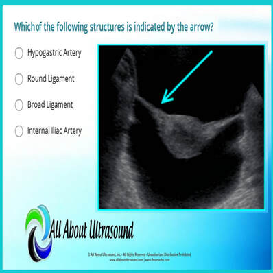
Which of the following structures is indicated by the arrow? ​Test your ultrasound anatomy skills! 

You guessed it! This image is referring to the Broad Ligament. 

The broad ligament is a two-layered fold of peritoneum that extends from the sides of the uterus to the floor and lateral walls of the pelvis inferiorly and the adnexa superiorly. The broad ligament helps to hold the uterus in its anatomic position. It covers the uterus, ovaries, and fallopian tubes and also includes nerves and blood vessels to these organs.

In this ultrasound image, the reason the broad ligament is easily identified is because of the fluid filling the pelvis. This highlights the location of the broad ligament. In normal settings, without a large amount of free fluid or the presence of other pathology, the broad ligament is rarely recognized on ultrasound. 

Studying for your OB/GYN ultrasound registry exam? Study with us! 
LEARN MORE

Share

0 Comments


Leave a Reply.

Details
    Picture

    Making Waves​™

    All About Ultrasound presents Making Waves™, our ultrasound specific blog and newsletter. Join us here for ultrasound news, cases and more! 

    Suggestions or Comments?
    Tell us your opinion...
    [email protected]

    Don't FORGET YOUR MERCH!

    SHOP NOW
    Picture

    Archives

    May 2023
    April 2023
    November 2022
    July 2020
    June 2020
    January 2019
    December 2018
    February 2018
    January 2018
    July 2017
    June 2017
    May 2017
    April 2017
    February 2017
    January 2017
    September 2016
    August 2016
    July 2016
    June 2016
    May 2016
    May 2015
    April 2015
    March 2015
    January 2015
    July 2014
    July 2013
    March 2013
    February 2013
    November 2012
    April 2012
    April 2011
    March 2011
    January 2011
    November 2010

    Categories

    All
    Accreditation
    Case Studies
    Echocardiography
    General
    Job Search
    OB/GYN
    Physics
    Registries
    Salary
    Scanning Tips
    Ultrasound News
    Ultrasound Research
    Vascular
    Webinar

    RSS Feed


ALL RIGHTS RESERVED
​​©ALL ABOUT ULTRASOUND, INC. dba iHEARTECHO

TERMS OF SERVICE   ●   PRIVACY POLICY   ●   
SUPPORT
​
[email protected]

ARDMS® is a registered trademark of the American Registry for Diagnostic Medical Sonography and is not associated with this website.  
CCI® is a registered trademark of Cardiovascular Credentialing International and is not associated with this website.
​
​WEB DESIGN BY: SoundMedia
  • Home
  • Ultrasound Services
    • Ultrasound Accreditation
    • Competency Assessments
  • Ultrasound Education
    • E-Learning >
      • E-Learning & CME
      • Upper Extremity Duplex
      • Venous Insufficiency
      • Doppler Principles and Hemodynamics
      • Left Ventricular Diastology
      • Constrictive Pericarditis
      • Aortic Stenosis
    • Free Membership
    • On-Site Training
    • Scanning Quick Guides
  • Ultrasound Registry Review
    • Free Membership
    • Upcoming Events
    • Ultrasound Physics SPI
    • Advanced Cardiac Sonographer
    • Adult Echocardiography
    • Pediatric Echocardiography
    • Fetal Echocardiography
    • Vascular Ultrasound
    • Abdomen Ultrasound
    • OB-GYN Ultrasound
    • Breast Ultrasound
    • Group | Team Access
  • Ultrasound Jobs
  • Ultrasound Resources
    • Ultrasound Case Studies
    • Sonography Coloring Book
    • Ultrasound Worksheets
    • Ultrasound Protocols
    • Ultrasound Credentialing
    • ULTRASOUND SCHOOLS
    • Sonography News
  • Making Waves™ Blog
  • SONO SHOP
  • Newsletter
  • FAQ
  • About Us
  • Contact Us
  • FREE SIGN UP
  • LOGIN
  • iHeartEcho