ULTRASOUND REGISTRY REVIEW SIMPLIFIED
Picture

Making Waves - All About Ultrasound BloG

  • Home
  • Ultrasound Registry Review
    • Ultrasound Physics SPI
    • Advanced Cardiac Sonographer
    • Adult Echocardiography
    • Pediatric Echocardiography
    • Fetal Echocardiography
    • Vascular Ultrasound
    • Abdomen Ultrasound
    • OB-GYN Ultrasound
    • Breast Ultrasound
    • E-Book Quiz
  • E-Learning & CME
  • Ultrasound Services
    • Ultrasound Education
    • Ultrasound Training
    • Ultrasound Accreditation
  • Ultrasound Resources
    • Ultrasound Excellence Award
    • Sonography Coloring Book
    • Ultrasound Worksheets
    • Ultrasound Protocols
    • Ultrasound Case Studies
    • Scanning Quick Guides
    • Ultrasound Credentialing
    • ULTRASOUND SCHOOLS
    • Sonography News
  • Blog
  • About Us
    • Newsletter
    • FAQ
    • Contact Us
    • iHeartEcho
  • SHOP
  • LOGIN

5/31/2023

Understanding Duodenal Atresia

0 Comments

Read Now
 
Understanding Duodenal Atresia: Causes, Symptoms, and Treatment

Duodenal atresia is a rare congenital condition that affects the development of the duodenum, the first part of the small intestine. It is a congenital intestinal obstruction awhich occurs when the duodenum is either completely blocked or narrowed, leading to problems with digestion and nutrient absorption. Let's talk about the causes, symptoms, and treatment options for duodenal atresia.

Causes:
Duodenal atresia is believed to be a result of abnormal development during the early stages of fetal growth. While the exact cause is unknown, several factors may contribute to its occurrence. These include genetic abnormalities, maternal diabetes, certain genetic syndromes such as Down syndrome, and exposure to certain medications during pregnancy.

Symptoms:
Duodenal atresia typically becomes apparent soon after birth. Some common symptoms after birth include:

  • Vomiting: Infants with duodenal atresia often experience vomiting, which can be bile-stained. This occurs due to the blockage preventing the passage of stomach contents into the small intestine.
  • Abdominal distention: The presence of a blockage in the duodenum can cause the abdomen to become swollen and distended.
  • Failure to thrive: Infants may have difficulty gaining weight and growing at a normal rate due to problems with digestion and nutrient absorption.
  • Dehydration: Vomiting can lead to dehydration if fluids are not adequately replaced.


Diagnosis:
Duodenal atresia is typically diagnosed shortly after birth. However, it can be identified on prenatal ultrasound. Ultrasound findings include:


  • Dilation of the stomach: One of the primary indicators of duodenal atresia is significant dilation of the fetal stomach. This occurs because the blockage in the duodenum prevents the passage of swallowed amniotic fluid to progress further along the digestive tract and the stomach becomes dilated with fluid.
  • Polyhydramnios: Polyhydramnios is commonly observed in cases of duodenal atresia. The lack of fetal swallowing due to the duodenal obstruction leads to reduced absorption of amniotic fluid into the intestine, resulting in its accumulation.
  • "Double-bubble" sign: The "double-bubble" sign is a classic ultrasound finding in duodenal atresia. It refers to the appearance of two distinct fluid-filled structures on the ultrasound image. The first bubble represents the dilated stomach, while the second bubble corresponds to the dilated portion of the duodenum just beyond the obstruction. This sign is highly suggestive of duodenal atresia.​
Picture


  • Dilated proximal small bowel loops: Due to the blockage at the level of the duodenum, the small bowel loops proximal to the obstruction may become dilated. These dilated loops can be visualized on ultrasound as fluid-filled structures that are larger than expected for the gestational age of the fetus.
  • Absence of fluid in the distal small bowel and colon: As a result of the duodenal obstruction, the fluid fails to pass through the duodenum and reach the distal small bowel and colon. This can be detected on ultrasound as a lack of fluid-filled structures in the lower abdomen.
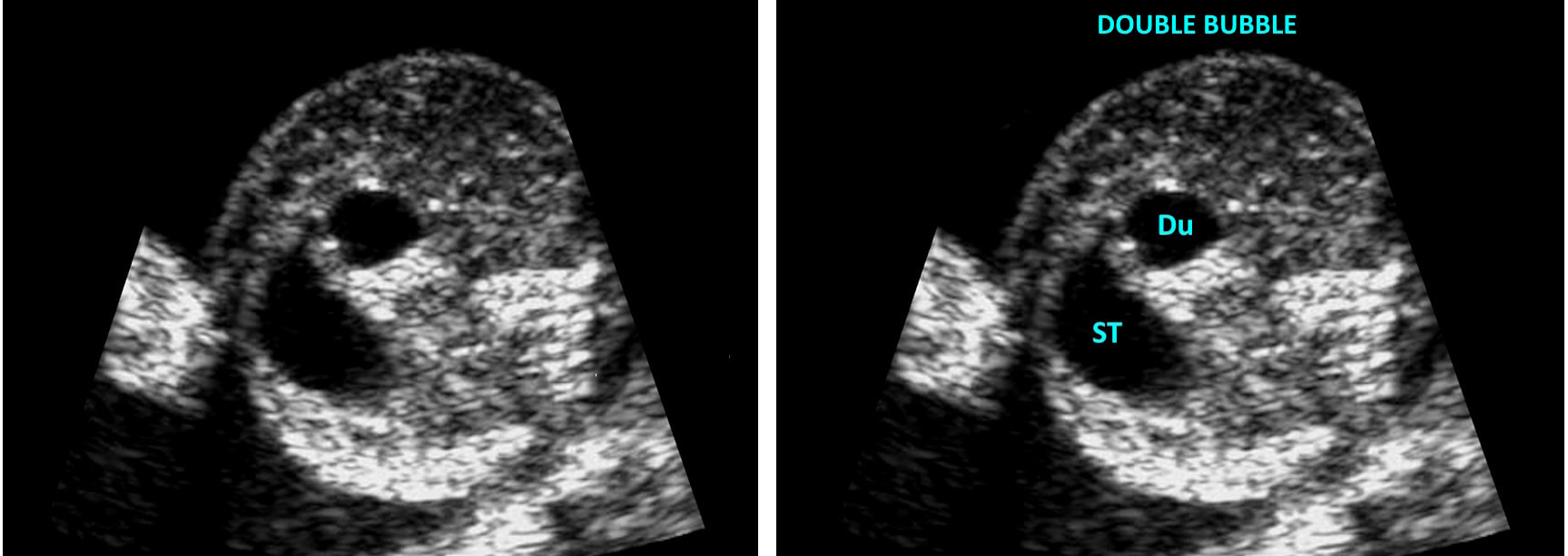
It is important to note that these ultrasound findings are suggestive of duodenal atresia, but they are not definitive. Additional diagnostic tests, such as genetic testing and fetal karyotyping, may be required for a confirmed diagnosis. Also, neonatal testing such as ultrasound and x-ray imaging can be helpful to diagnose and evaluate the severity.

Treatment:
The primary treatment for duodenal atresia is surgery. The surgical procedure involves bypassing or removing the obstructed portion of the duodenum and connecting the healthy segments. The specific surgical approach depends on the severity of the condition.

  • Open surgery: In some cases, open surgery may be required to access and repair the blockage in the duodenum.
  • Laparoscopic surgery: Minimally invasive laparoscopic techniques may be used for less severe cases, where smaller incisions are made, reducing the recovery time and scarring.

Following surgery, infants will require close monitoring in a neonatal intensive care unit (NICU) to ensure their digestive system functions properly. They may receive nutrition through intravenous fluids until they are able to tolerate oral feeding.

Prognosis:
With timely diagnosis and appropriate surgical intervention, the outlook for infants with duodenal atresia is generally favorable. After surgery, most infants can resume normal feeding and achieve healthy growth and development. However, it is essential for parents and caregivers to follow up with regular medical check-ups to monitor the child's progress and ensure there are no long-term complications related to the surgery. With proper treatment and ongoing medical care, children with duodenal atresia can go on to lead healthy and fulfilling lives. 

Share

0 Comments


Leave a Reply.

Details
    Picture

    Making Waves

    All About Ultrasound presents Making Waves, our ultrasound specific blog. Join us here for ultrasound news, cases and more! 

    Suggestions or Comments?
    Tell us your opinion...
    admin@allaboutultrasound.com

    Don't FORGET YOUR MERCH!

    SHOP NOW
    Picture

    Archives

    May 2023
    April 2023
    November 2022
    July 2020
    June 2020
    January 2019
    December 2018
    February 2018
    January 2018
    July 2017
    June 2017
    May 2017
    April 2017
    February 2017
    January 2017
    September 2016
    August 2016
    July 2016
    June 2016
    May 2016
    May 2015
    April 2015
    March 2015
    January 2015
    July 2014
    July 2013
    March 2013
    February 2013
    November 2012
    April 2012
    April 2011
    March 2011
    January 2011
    November 2010

    Categories

    All
    Accreditation
    Case Studies
    Echocardiography
    General
    Job Search
    OB/GYN
    Physics
    Registries
    Salary
    Scanning Tips
    Ultrasound News
    Ultrasound Research
    Vascular
    Webinar

    RSS Feed


ALL RIGHTS RESERVED
​​©ALL ABOUT ULTRASOUND, INC. dba iHEARTECHO

TERMS OF SERVICE   ●   PRIVACY POLICY   ●   
SUPPORT
​
support@allaboutultrasound.com

ARDMS® is a registered trademark of the American Registry for Diagnostic Medical Sonography and is not associated with this website.  
CCI® is a registered trademark of Cardiovascular Credentialing International and is not associated with this website.
​
​WEB DESIGN BY: LAUNCH
  • Home
  • Ultrasound Registry Review
    • Ultrasound Physics SPI
    • Advanced Cardiac Sonographer
    • Adult Echocardiography
    • Pediatric Echocardiography
    • Fetal Echocardiography
    • Vascular Ultrasound
    • Abdomen Ultrasound
    • OB-GYN Ultrasound
    • Breast Ultrasound
    • E-Book Quiz
  • E-Learning & CME
  • Ultrasound Services
    • Ultrasound Education
    • Ultrasound Training
    • Ultrasound Accreditation
  • Ultrasound Resources
    • Ultrasound Excellence Award
    • Sonography Coloring Book
    • Ultrasound Worksheets
    • Ultrasound Protocols
    • Ultrasound Case Studies
    • Scanning Quick Guides
    • Ultrasound Credentialing
    • ULTRASOUND SCHOOLS
    • Sonography News
  • Blog
  • About Us
    • Newsletter
    • FAQ
    • Contact Us
    • iHeartEcho
  • SHOP
  • LOGIN