
|
It is so easy to get caught up with our patients who need our immediate attention for things that aren't even ultrasound related and even seasoned sonographers can ignore physics altogether, but when we take it back to the basics, this is where image optimization begins. So, let's take a step back and look at the basics again..... Doppler principles and hemodynamics... I know, I know, for some just saying the words brings tears to their eyes as they recall sleepless nights studying for the Ultrasound Physics registry! But no need to fear! Doppler is simple when you break it down. So the Doppler Effect is quite simply either a positive shift which is a compression in the wavelength (a higher frequency) or a negative shift, which results in an elongated wavelength, (a lower frequency) and this is of course all relative to the observer. 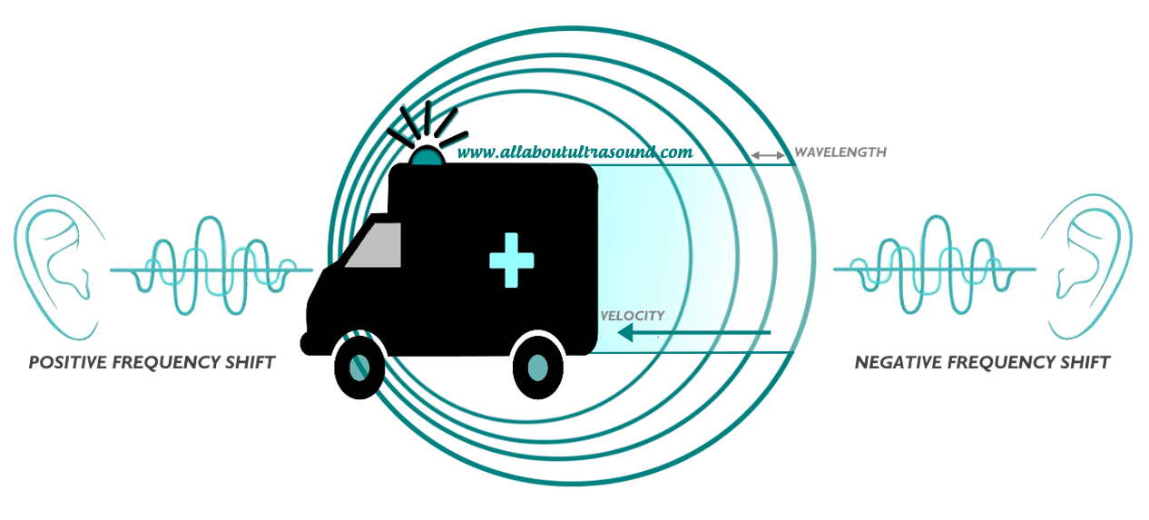 So what does this have to do with Doppler ultrasound? I'm glad you asked. Doppler frequency shifts come from moving red blood cells and give us the spectral display that we see in Continuous Wave and Pulse Wave Doppler. The velocities obtained from these frequency shifts can also be displayed in Color Doppler, superimposed on top of the 2D ultrasound image. This is a method of displaying the mean or average velocity, while the spectral Doppler display can quantify specific velocities, like the peak systolic and end diastolic velocities, at a point in time. Because the Doppler frequency shift is relative to the position of the observer, this is important when placing the Doppler sample and angle of insonation. The Doppler sample volume is the "observer" and the frequency shift created by the moving red blood cell is what we are evaluating on the spectral display. But what happens if the observer changes position? The velocity observed will be different. This is extremely important for Doppler Angle. The Society for Vascular Ultrasound recommends that scanning angle be maintained between 45-60 degrees. While we know that the closer to 0 degrees, the more accurate the velocity result because of the math and the cosine of the angle, try getting that on a your average carotid Doppler. It is often not attainable and so for reporting and to maintain consistency in the lab, it is best to stay within the range of what can be easily obtained on most exams. 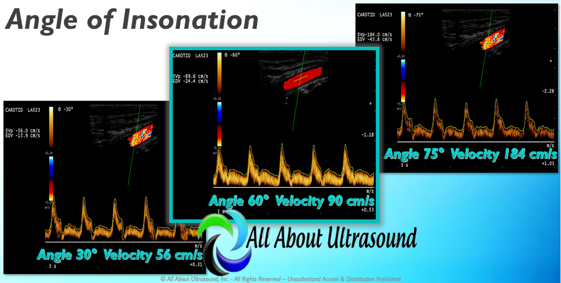 So as you can see from the image, as the angle of insonation is moved, the velocity result is SIGNIFICANTLY impacted. This can be a very big factor in following up serial ultrasound studies if the same Doppler angle is not used for follow up exams. This is why it is so important to look at the previous study images, especially if there is disease. So when you're scanning don't forget these very basic settings and factors that can have a large impact on your patient's results. Studying for your SPI ULTRASOUND PHYSICS REGISTRY? We have you covered with our SPI Ultrasound Physics Registry Review course and FREE TRIAL available! Want more info on Doppler Principles and Hemodynamics? Need CME's?
See our Mastering Doppler Principles and Hemodynamics E-Learning CME Course! Approved for 1 SDMS CME Credit Hour
5 Comments
Patience
12/19/2018 10:06:03 am
Thanks. Quite simplified.
Yvonne Valov-Celso RVT
3/19/2019 08:22:43 am
Well written
Brenda
12/19/2018 11:22:22 pm
I need a better understanding of UI Leave a Reply. |

Making Waves™All About Ultrasound presents Making Waves™, our ultrasound specific blog and newsletter. Join us here for ultrasound news, cases and more! Don't FORGET YOUR MERCH!
Archives
May 2023
Categories
All
|
 RSS Feed