
|
How would you adjust your ultrasound settings to go about proving this diagnosis? What waveform characteristics would you be aware of in adjacent vasculature?   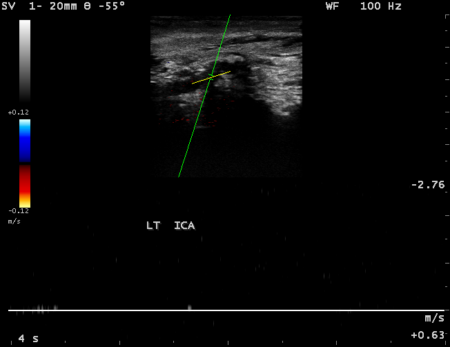 Images courtesy of SONA IMAGING SOLUTIONS
3 Comments
Tammy
3/23/2011 03:07:07 am
Is this an occluded ICA? Have to watch waveforms for internalization of the ECA, as it may take on characteristics of the ICA waveform.
Jose
6/12/2011 12:09:11 pm
You will have to decrease the color PRF ( scale ) to increase sensitivity to the slowest possible flow that might exists in that ICA. Also A high resitance doppler pattern in the more proximal segment (CCA) would be suggestive of an ICA occlusion. External to internal collateralization by obtaining a low resistance flow pattern in the ECA will confirm the ICA occlusion ( in this case the ipsilateral ophthalmic artery will have reversal of the flow. Also a contralateral ICA increase of the flow will compensate for that ICA occlusion, especially if there is an Anterior to Anterior collateralization. Leave a Reply. |

Making Waves™All About Ultrasound presents Making Waves™, our ultrasound specific blog and newsletter. Join us here for ultrasound news, cases and more! Don't FORGET YOUR MERCH!
Archives
May 2023
Categories
All
|
 RSS Feed