ULTRASOUND REGISTRY REVIEW SIMPLIFIED
Picture

Making Waves - All About Ultrasound BloG

  • Home
  • Ultrasound Registry Review
    • Ultrasound Physics SPI
    • Advanced Cardiac Sonography
    • Adult Echocardiography
    • Pediatric Echocardiography
    • Fetal Echocardiography
    • Vascular Ultrasound
    • Abdomen Ultrasound
    • OB-GYN Ultrasound
    • Breast Ultrasound
    • E-Book Quiz
  • E-Learning & CME
  • Ultrasound Services
    • LIVE Online Tutoring
    • Ultrasound Training
  • Ultrasound Resources
    • Ultrasound Excellence Award
    • Sonography Coloring Book
    • Ultrasound Worksheets
    • Ultrasound Protocols
    • Ultrasound Case Studies
    • Scanning Quick Guides
    • Ultrasound Credentialing
    • ULTRASOUND SCHOOLS
    • Sonography News
  • Blog
  • About Us
    • Newsletter
    • FAQ
    • Contact Us
    • iHeartEcho
  • SHOP
  • LOGIN

1/9/2019

7 Study Tips to Pass your Ultrasound Registry Exam

2 Comments

Read Now
 
Picture
Studying for that ARDMS or CCI ultrasound registry exam can be brutal!
So we've covered this topic a few times before, but it is always helpful to revisit some of these study tips. Passing your ARDMS or CCI Ultrasound Registry exam is easier than you think.... There's no need to worry!

Don't stress over it... just study. Mastering ultrasound physics or any other ultrasound specialty can be so overwhelming and often it can you make you want to give up. Don't give up! You've got this! Keep on studying and use these simple tips to study and PASS your ultrasound registry! 
  • ​1. Get Organized. Make a plan and a schedule for your study time. It helps if you are a new grad to begin while you are still in school. Know what you need to prepare in order to apply (that alone can be a daunting task). Also check out the ARDMS website and review the content outline for the exam you are taking. Then make studying a priority. Find the best time and schedule that works for and stick to it, no matter what! No procrastinating! (I'm preaching to the choir on this one).
  • 2. Don't multitask! Our lives are so hectic and most of the time, our lives require multitasking, but surprisingly studies have shown that multitasking is physically impossible. Multitasking actually keeps you from focusing on what you need to master. So, master the basics and stay focused on one thing at a time.
  • 3. Split it up! Studying usually isn't fun and forcing your way through a long study marathon will only make it worse. The saying "you eat an elephant one bite a time",  applies here. Divide your work into manageable chunks. Take one section and master that! Reward yourself when you finish each accomplishment to keep it fun and less stressful! 
  • 4. Sleep - Sleep is so important! Getting those eight hours of zzz's every night will keep your focus sharp and improve your working memory. Don't skip your sleep - get your rest! (again preaching to the choir here)
  • 5. Take notes - Taking notes will not only keep you more engaged during class or any review courses, but will also help you narrow down what you need to study when exam time rolls around. Also, take notes in pencil.... the tactile engagement of the pencil will help you remember the content when you need it! 
  • 6. Work with what you have - Engage as many senses in studying as you can... read, write and speak the content. Have someone else read the content back to you and quiz yourself. This will help you remember the content on the big day!
  • 7. Study! Obvious, right? There's a right and a wrong way to study... get it right! Review the material several days ahead of time. Look over it in small chunks, and in different ways (for example, write flashcards one day and take practice tests the next).  And.... Don't cram. Give yourself time!

Hope these tips help you pass your ultrasound registry. For more help and tools to pass - check out our ultrasound registry reviews with quizzes and tools to help you master the ARDMS and CCI registries! Sign up for a FREE TRIAL (no credit card needed) of our Ultrasound Physics SPI Registry Review Course that includes our PASS GUARANTEE and pass with us!


Picture

Share

2 Comments

7/29/2017

Step by Step Guide to Mesenteric Abdominal Duplex

5 Comments

Read Now
 
Picture
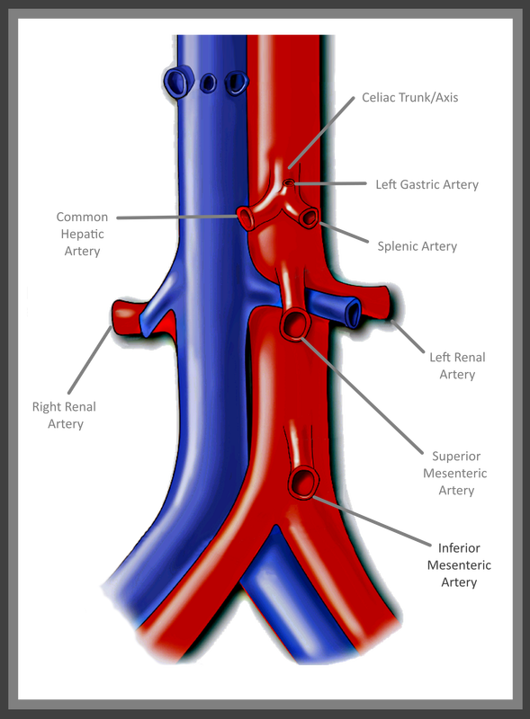
Mesenteric Abdominal Duplex – it sounds complicated right? In reality though, as long as your patient is prepped, there’s not much to it. I say that with caution though – we’ve all had those patients that just really need another modality of testing. Let's be real... we all know that our ultrasound transducer is NOT a magic wand! 

First and foremost - Patient Prep! Your patient prep is one of the most important factors when performing Mesenteric Duplex Ultrasound. Your patient needs to be NPO for at least 6-8 hours before scanning. A test done without the right prep, might as well have not been done at all.  This is for a number of reasons:
1. You can't see squat with air and gas in the way!
2. The mesenteric arterial system should be scanned both pre and post prandial for evaluation of stenosis and arterial ischemic response. ​

Picture
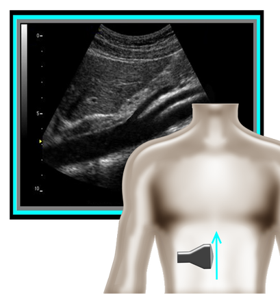
​So where to begin and what protocol? I've got that covered for you! A mesenteric duplex protocol should include, at a minimum...  Transverse and Longitudinal approach with 2D, Longitudinal approach with Color and PW Doppler (Record PSV and EDV) at each of the following locations:                                                                                                                                                       
•Proximal Aorta
•Celiac Artery/Trunk
•Branching of Common Hepatic Artery and Splenic Artery from the Celiac Artery
•SMA origin, proximal, mid, distal
•IMA origin, proximal and as distal as possible
•IVC 

​A few things can help you improve your imaging and will also help you evaluate patient pathology.
Picture
Picture
1. Patient position is an important factor to remember. You should position your patient in a slightly elevated/supine position. This allows for a better view with less pressure on the diaphragm. Another trick is to have your patient lay supine and bend their knees. This also reduces pressure and tension on the diaphragm and allows for better visualization of the structures near the xyphoid process (ie. proximal aorta, celiac trunk, SMA). Having the patient sit erect can also help with evaluation of median arcuate ligament compression and if images are limited otherwise.
​
​2. Using patient respirations to control the movement of structures and improve image quality (especially for Doppler waveforms). Respirations also help to evaluate for median arcuate ligament compression is sometimes a major factor in diagnosing this condition. Patient's with median arcuate ligament compression will have mildly elevated flow in the Celiac Artery with inspiration (this is normal). However, with expiration, the artery will be compressed by the median arcuate ligament and will cause an extrinsic obstruction due to compression and a marked increase in flow velocity. Using patient respirations to assess for this is a major factor of the Mesenteric Duplex exam. 

​3. Use Power Doppler - on those difficult patients, power Doppler can be your savior! This can also be used to visualize the Celiac Branches, the IMA and other smaller vessels.
Picture
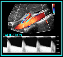
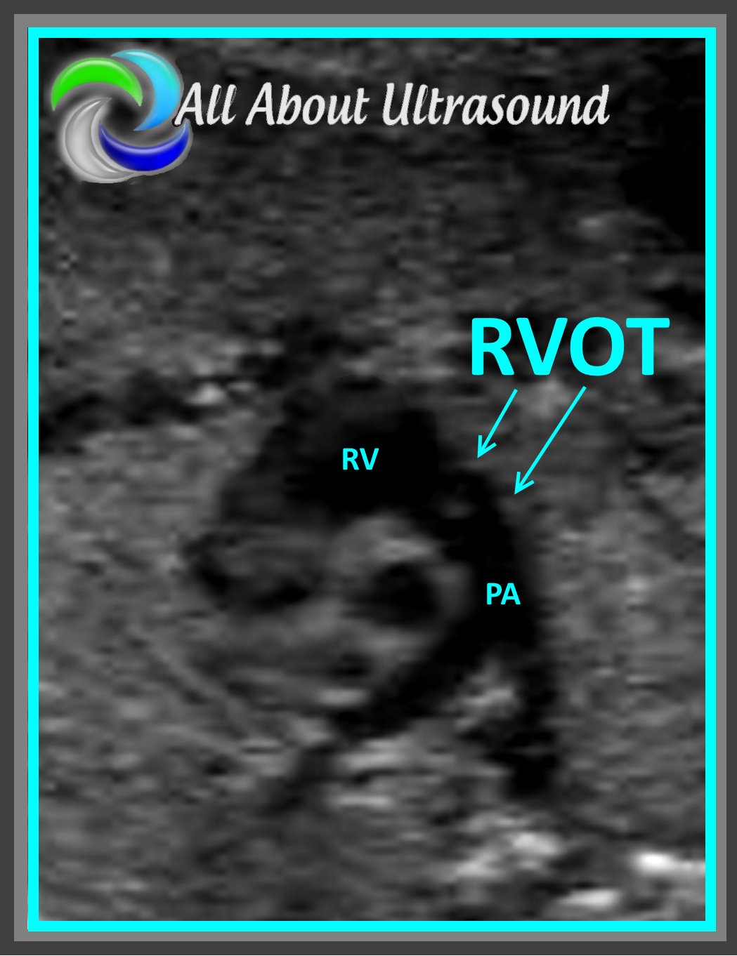
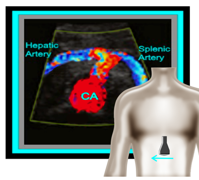
Okay, so now that we have the basics out of the way... you're probably wondering, what should the waveforms look like and what is normal/abnormal? So here's a quick guide:

​Celiac Artery
​

Remember that the Celiac Artery supplies the liver, spleen and stomach, which are low resistance vascular beds. Normal Doppler waveforms will show increased diastolic flow because of the organs supplied. Flow may also increase with inspiration. ​

Don't forget to evaluate the branches! This is best done in a transverse plane. 

≥70% Celiac Artery Stenosis will show a peak systolic velocity of  ≥200 cm/s.
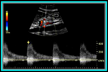
Picture
​Superior Mesenteric Artery
​

​Remember that the SMA supplies the jejunum, ileum, and both the right and transverse colon. Because of this, waveform characteristics will vary based on state (ie. NPO, post-prandial). Diastolic flow will increase as needed for digestion, post-prandially.

In the normal vessel, post-prandial evaluation should show increased peak systolic flow velocities. If PSV flow does not increase, this is suggestive of a hemodynamically significant stenosis. Also keep in mind that inspiration will show an increase in peak systolic velocities.

≥70% SMA stenosis will show a peak systolic velocity of  ≥275cm/s or absence of color flow in the SMA. End diastolic flow velocities of ≥45cm/s are also an indication of ≥70% SMA stenosis. 

​Also keep in mind that you can find pathology based on the angle of the SMA  takeoff from the aorta. If the angle is markedly increased, it may indicate the presence of adenopathy. The SMA should course parallel to the aorta. 


Inferior Mesenteric Artery

​The IMA supplies the distal 1/3 of the transverse colon, splenic flexure, descending colon, sigmoid colon and rectum. It's waveform characteristics are similar to the SMA and will vary based on state (ie. NPO, post-prandial). Diastolic flow will increase as needed for digestion, post-prandially.

≥70% IMA stenosis will show a peak systolic velocity of  ≥275cm/s or absence of color flow in the IMA.

​You may need to get your magic wand out for this one! 
Picture
Diagnostic Criteria Reference: Moneta, et al

GET OUR FREE SCANNING TIPS QUICK GUIDE

FREE DOWNLOAD
Picture

Share

5 Comments

7/20/2017

5 Best Practice Tips to Ob-Gyn Ultrasound

16 Comments

Read Now
 
1. That Bladder! We have all seen the pregnant pee pee dance… While sometimes uncomfortable for our patients, this is the key to the first trimester transabdominal approach and also to 2nd trimester cervical measurements. Sometimes it just can’t be avoided, your patient is going to dance! However, in the 2nd trimester, it can be helpful for your patient to get what you need with the bladder full and to let them empty it out before completing the rest of your study. Often this can also improve the position of the fetus and help you to get better images with a relaxed mama and a relaxed baby!
Picture
Picture
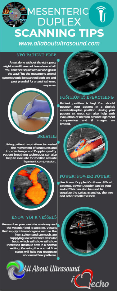
2. Scanning and trying to find those ovaries can prove to be a challenge with a pregnant uterus! Even though it is not our focus, we can’t forget those ovaries! Making sure to image those along with the uterus and fetal images will ensure that you’re not missing potential pathology, but it can definitely be tough with that pregnant uterus in the way. Best methods are to bring the transducer laterally to the patient’s side and to scan in a transverse plane along the lateral side of the uterus, starting pretty high up and moving down toward the patient’s cervix. That transverse plane will give you an increased field of view and will allow you to identify structures more easily.

3. Outflow tracts… the sound of that might scare some sonographers who are not used to looking at the fetal heart or evaluating this on a regular basis, but it’s much easier than you might think. Remember there are two main blood vessels that exit the heart – the aorta and the pulmonary artery. That’s it… just two. So how do you tell which is which? Most of the time (unless in a case of transposition of the great arteries or double outlet right ventricle), the aorta exits the left ventricle and the pulmonary artery exits the right ventricle. It’s important to note that the aorta will arch around after it leaves the heart and extend into the thoracic/abdominal aorta. The pulmonary artery will typically split into two branches.

​Yes, there are many variations that can happen with cardiac pathology and unless you specialize in maternal fetal medicine or fetal echocardiography, you may not know the specifics of every congenital heart disease you might encounter, but if you know the basics and what that looks like, you should be able to know when it’s just not right and alert your reading physician. If you’re not scanning this on a regular basis, it would be a good idea to add this to your daily protocol.
Picture
Picture
Picture
4. Amniotic Fluid Index – how do you know your measurements are accurate? Many sonographers are not aware of how to properly measure fluid for an AFI. One key tip is to scan with color Doppler flow on – this allows you to identify umbilical cord that might be floating within the pocket of fluid. Another best practice tip and proper way to perform AFI measurements is to be sure your patient is lying flat on the table and keep the probe/ultrasound beam perpendicular to the floor. Also, keep your measurements perpendicular as well. This helps to ensure that the fluid pocket is not falsely enlarged.
5. Gender! We’ve all been there, where it seems that everyone is there for the show. While somehow you ended up being the entertainment for the expectant parents, don’t forget how exciting it can be to know how to plan for your little one. Yes, it’s often a pain with the dad, grandma, grandpa, sister, brother, niece, nephew, the mailman, the neighbor, the sister’s boyfriend’s mother-in-law and everyone else trying to squeeze into the room to catch a glance of the precious cargo and whatever package the baby might be sporting… so take a deep breath, don’t lose it on grandma and remember to SMILE 😊. Oh and scanning, well you know how that goes…​ get those pesky diagnostic measurements and anatomy evaluations out of the way and  then... turtle or hamburger… take your pick!
Picture
Picture

​

Share

16 Comments

3/23/2011

Ultrasound Accreditation

3 Comments

Read Now
 
Ultrasound Accreditation and/or sonographer credentialing requirements are already in effect in many states and soon will be required for reimbursement of exams. Is your ultrasound lab accredited? If not, are the sonographers in your department registered? How is your lab preparing for the upcoming accreditation requirements?

Share

3 Comments

11/8/2010

Our new blog!

3 Comments

Read Now
 
Picture
Thank you for visiting All About Ultrasound. We are making changes here to bring you the sonography resources that you need. We are dedicated to making resources readily available to the ultrasound community. We have adapted our previously for fee advertising to allow for a free service in order to get you the job postings, cme credits and other resources you need right into your hands. Check out our newly updated website and don't forget to share us with your colleagues and friends!

Share

3 Comments
Details
    Picture

    Making Waves

    All About Ultrasound presents Making Waves, our ultrasound specific blog. Join us here for ultrasound news, cases and more! 

    Suggestions or Comments?
    Tell us your opinion...
    admin@allaboutultrasound.com

    Don't FORGET YOUR MERCH!

    SHOP NOW
    Picture

    Archives

    November 2022
    July 2020
    June 2020
    January 2019
    December 2018
    February 2018
    January 2018
    July 2017
    June 2017
    May 2017
    April 2017
    February 2017
    January 2017
    September 2016
    August 2016
    July 2016
    June 2016
    May 2016
    May 2015
    April 2015
    March 2015
    January 2015
    July 2014
    July 2013
    March 2013
    February 2013
    November 2012
    April 2012
    April 2011
    March 2011
    January 2011
    November 2010

    Categories

    All
    Accreditation
    Case Studies
    Echocardiography
    General
    Job Search
    OB/GYN
    Physics
    Registries
    Salary
    Scanning Tips
    Ultrasound News
    Ultrasound Research
    Vascular
    Webinar

    RSS Feed


ALL RIGHTS RESERVED
​​©ALL ABOUT ULTRASOUND, INC. dba iHEARTECHO

TERMS OF SERVICE   ●   PRIVACY POLICY   ●   
SUPPORT
​
support@allaboutultrasound.com

ARDMS® is a registered trademark of the American Registry for Diagnostic Medical Sonography and is not associated with this website.  
CCI® is a registered trademark of Cardiovascular Credentialing International and is not associated with this website.
​
​WEB DESIGN BY: LAUNCH
  • Home
  • Ultrasound Registry Review
    • Ultrasound Physics SPI
    • Advanced Cardiac Sonography
    • Adult Echocardiography
    • Pediatric Echocardiography
    • Fetal Echocardiography
    • Vascular Ultrasound
    • Abdomen Ultrasound
    • OB-GYN Ultrasound
    • Breast Ultrasound
    • E-Book Quiz
  • E-Learning & CME
  • Ultrasound Services
    • LIVE Online Tutoring
    • Ultrasound Training
  • Ultrasound Resources
    • Ultrasound Excellence Award
    • Sonography Coloring Book
    • Ultrasound Worksheets
    • Ultrasound Protocols
    • Ultrasound Case Studies
    • Scanning Quick Guides
    • Ultrasound Credentialing
    • ULTRASOUND SCHOOLS
    • Sonography News
  • Blog
  • About Us
    • Newsletter
    • FAQ
    • Contact Us
    • iHeartEcho
  • SHOP
  • LOGIN