ULTRASOUND REGISTRY REVIEW SIMPLIFIED
Picture
   

Making Waves™ - All About Ultrasound BloG

  • Home
  • Ultrasound Services
    • Ultrasound Accreditation
    • Competency Assessments
  • Ultrasound Education
    • E-Learning >
      • E-Learning & CME
      • Upper Extremity Duplex
      • Venous Insufficiency
      • Doppler Principles and Hemodynamics
      • Left Ventricular Diastology
      • Constrictive Pericarditis
      • Aortic Stenosis
    • Free Membership
    • On-Site Training
    • Scanning Quick Guides
  • Ultrasound Registry Review
    • Free Membership
    • Upcoming Events
    • Ultrasound Physics SPI
    • Advanced Cardiac Sonographer
    • Adult Echocardiography
    • Pediatric Echocardiography
    • Fetal Echocardiography
    • Vascular Ultrasound
    • Abdomen Ultrasound
    • OB-GYN Ultrasound
    • Breast Ultrasound
    • Group | Team Access
  • Ultrasound Jobs
  • Ultrasound Resources
    • Ultrasound Case Studies
    • Sonography Coloring Book
    • Ultrasound Worksheets
    • Ultrasound Protocols
    • Ultrasound Credentialing
    • ULTRASOUND SCHOOLS
    • Sonography News
  • Making Waves™ Blog
  • SONO SHOP
  • Newsletter
  • FAQ
  • About Us
  • Contact Us
  • FREE SIGN UP
  • LOGIN
  • iHeartEcho

12/19/2018

Mastering Doppler Principles and Hemodynamics

5 Comments

Read Now
 
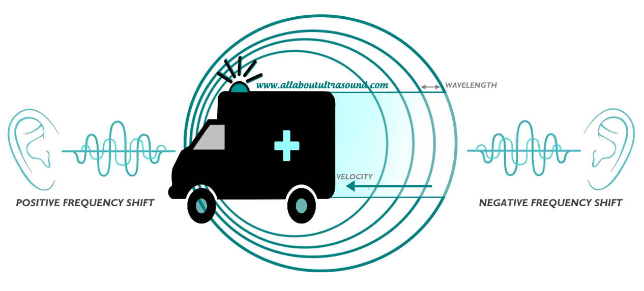
It is so easy to get caught up with our patients who need our immediate attention for things that aren't even ultrasound related and even seasoned sonographers can ignore physics altogether, but when we take it back to the basics, this is where image optimization begins. So, let's take a step back and look at the basics again.....  Doppler principles and hemodynamics... I know, I know, for some just saying the words brings tears to their eyes as they recall sleepless nights studying for the Ultrasound Physics registry! But no need to fear! Doppler is simple when you break it down. So the Doppler Effect is quite simply either a positive shift which is a compression in the wavelength (a higher frequency)  or a negative shift, which results in an elongated wavelength, (a lower frequency) and this is of course all relative to the observer. 
Picture
So what does this have to do with Doppler ultrasound? I'm glad you asked.
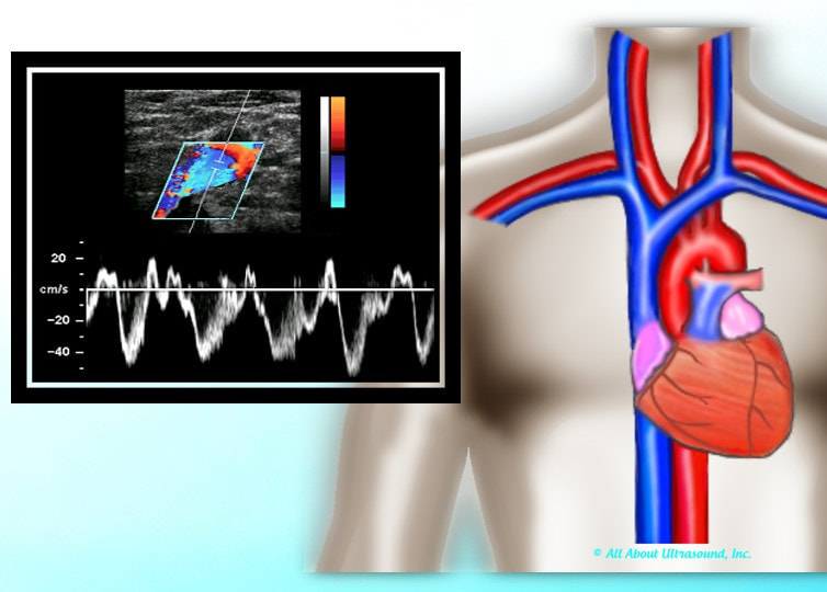
​Doppler frequency shifts come from moving red blood cells and give us the spectral display that we see in Continuous Wave and Pulse Wave Doppler. The velocities obtained from these frequency shifts can also be displayed in Color Doppler, superimposed on top of the 2D ultrasound image.  This is a method of displaying the mean or average velocity, while the spectral Doppler display can quantify specific velocities, like the peak systolic and end diastolic velocities, at a point in time. 

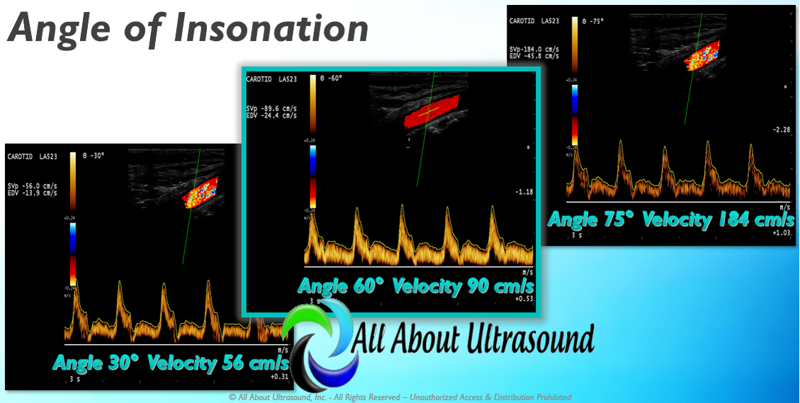
Because the Doppler frequency shift is relative to the position of the observer, this is important when placing the Doppler sample and angle of insonation. The Doppler sample volume is the "observer" and the frequency shift created by the moving red blood cell is what we are evaluating on the spectral display. But what happens if the observer changes position? The velocity observed will be different.  This is extremely important for Doppler Angle. The Society for Vascular Ultrasound recommends that scanning angle be maintained between 45-60 degrees.  While we know that the closer to 0 degrees, the more accurate the velocity result because of the math and the cosine of the angle, try getting that on a your average carotid Doppler. It is often not attainable and so for reporting and to maintain consistency in the lab, it is best to stay within the range of what can be easily obtained on most exams. 
Picture
So as you can see from the image, as the angle of insonation is moved, the velocity result is SIGNIFICANTLY impacted. This can be a very big factor in following up serial ultrasound studies if the same Doppler angle is not used for follow up exams. This is why it is so important to look at the previous study images, especially if there is disease. So when you're scanning don't forget these very basic settings and factors that can have a large impact on your patient's results. 
Studying for your SPI ULTRASOUND PHYSICS REGISTRY?
​We have you covered with our SPI Ultrasound Physics Registry Review course and FREE TRIAL available!

Picture
Want more info on Doppler Principles and Hemodynamics? Need CME's?
​See our Mastering Doppler Principles and Hemodynamics E-Learning CME Course!

Approved for 1 SDMS CME Credit Hour

Share

5 Comments

2/19/2018

Mastering Upper Extremity Duplex

1 Comment

Read Now
 
Master the concepts of Upper Extremity Duplex, including Hemodialysis Access and More! Our new CME Opportunity with E-Learning On Demand. This E-Learning course has been approved for 2 SDMS CME Credit Hours. Register today! CLICK HERE
Picture
Register Now!

Share

1 Comment

1/1/2018

Pediatric Echo Registry Review

0 Comments

Read Now
 
It is application time again for the ARDMS® Pediatric Echo Registry. The next time to apply is January 3, 2018-March 6, 2018​. Apply for your registry and study with us today! Our Pediatric Echo Ultrasound Registry Review Quiz starts at just $19.99 and can help you prepare for that dreaded registry exam. No need to be intimidated. With over 200 questions in our unlimited quiz, our Test & Learn format will help you learn as you go, testing your knowledge and teaching too! Our test responses are based on your answers and questions are designed to help you learn as you go. Don't be overwhelmed by ultrasound physics or any other ultrasound specialty registry exam.... we have the tools to help you master the content and pass your ARDMS/CCI registries. Simple. Easy. Done! ​
Share

0 Comments

7/29/2017

Step by Step Guide to Mesenteric Abdominal Duplex

6 Comments

Read Now
 
Picture
Mesenteric Abdominal Duplex – it sounds complicated right? In reality though, as long as your patient is prepped, there’s not much to it. I say that with caution though – we’ve all had those patients that just really need another modality of testing. Let's be real... we all know that our ultrasound transducer is NOT a magic wand! 

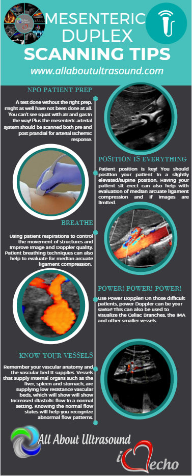
First and foremost - Patient Prep! Your patient prep is one of the most important factors when performing Mesenteric Duplex Ultrasound. Your patient needs to be NPO for at least 6-8 hours before scanning. A test done without the right prep, might as well have not been done at all.  This is for a number of reasons:
1. You can't see squat with air and gas in the way!
2. The mesenteric arterial system should be scanned both pre and post prandial for evaluation of stenosis and arterial ischemic response. ​
Picture
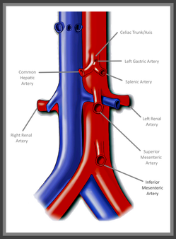
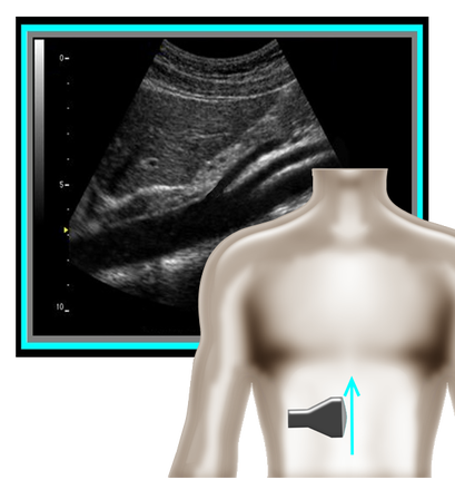
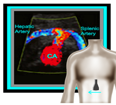
​So where to begin and what protocol? I've got that covered for you! A mesenteric duplex protocol should include, at a minimum...  Transverse and Longitudinal approach with 2D, Longitudinal approach with Color and PW Doppler (Record PSV and EDV) at each of the following locations:                                                                                                                                                       
•Proximal Aorta
•Celiac Artery/Trunk
•Branching of Common Hepatic Artery and Splenic Artery from the Celiac Artery
•SMA origin, proximal, mid, distal
•IMA origin, proximal and as distal as possible
•IVC 

​A few things can help you improve your imaging and will also help you evaluate patient pathology.
Picture
Picture
1. Patient position is an important factor to remember. You should position your patient in a slightly elevated/supine position. This allows for a better view with less pressure on the diaphragm. Another trick is to have your patient lay supine and bend their knees. This also reduces pressure and tension on the diaphragm and allows for better visualization of the structures near the xyphoid process (ie. proximal aorta, celiac trunk, SMA). Having the patient sit erect can also help with evaluation of median arcuate ligament compression and if images are limited otherwise.
​
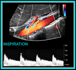
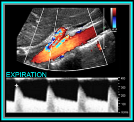
​2. Using patient respirations to control the movement of structures and improve image quality (especially for Doppler waveforms). Respirations also help to evaluate for median arcuate ligament compression is sometimes a major factor in diagnosing this condition. Patient's with median arcuate ligament compression will have mildly elevated flow in the Celiac Artery with inspiration (this is normal). However, with expiration, the artery will be compressed by the median arcuate ligament and will cause an extrinsic obstruction due to compression and a marked increase in flow velocity. Using patient respirations to assess for this is a major factor of the Mesenteric Duplex exam. 

​3. Use Power Doppler - on those difficult patients, power Doppler can be your savior! This can also be used to visualize the Celiac Branches, the IMA and other smaller vessels.
Picture
Okay, so now that we have the basics out of the way... you're probably wondering, what should the waveforms look like and what is normal/abnormal? So here's a quick guide:

​Celiac Artery
​

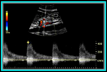
Remember that the Celiac Artery supplies the liver, spleen and stomach, which are low resistance vascular beds. Normal Doppler waveforms will show increased diastolic flow because of the organs supplied. Flow may also increase with inspiration. ​

Don't forget to evaluate the branches! This is best done in a transverse plane. 

≥70% Celiac Artery Stenosis will show a peak systolic velocity of  ≥200 cm/s.
Picture
​Superior Mesenteric Artery
​

​Remember that the SMA supplies the jejunum, ileum, and both the right and transverse colon. Because of this, waveform characteristics will vary based on state (ie. NPO, post-prandial). Diastolic flow will increase as needed for digestion, post-prandially.

In the normal vessel, post-prandial evaluation should show increased peak systolic flow velocities. If PSV flow does not increase, this is suggestive of a hemodynamically significant stenosis. Also keep in mind that inspiration will show an increase in peak systolic velocities.

≥70% SMA stenosis will show a peak systolic velocity of  ≥275cm/s or absence of color flow in the SMA. End diastolic flow velocities of ≥45cm/s are also an indication of ≥70% SMA stenosis. 

​Also keep in mind that you can find pathology based on the angle of the SMA  takeoff from the aorta. If the angle is markedly increased, it may indicate the presence of adenopathy. The SMA should course parallel to the aorta. 


Inferior Mesenteric Artery

​The IMA supplies the distal 1/3 of the transverse colon, splenic flexure, descending colon, sigmoid colon and rectum. It's waveform characteristics are similar to the SMA and will vary based on state (ie. NPO, post-prandial). Diastolic flow will increase as needed for digestion, post-prandially.

≥70% IMA stenosis will show a peak systolic velocity of  ≥275cm/s or absence of color flow in the IMA.

​You may need to get your magic wand out for this one! 
Picture
Diagnostic Criteria Reference: Moneta, et al

GET OUR FREE SCANNING TIPS QUICK GUIDE
FREE DOWNLOAD
Picture

Share

6 Comments

7/20/2017

5 Best Practice Tips to Ob-Gyn Ultrasound

16 Comments

Read Now
 
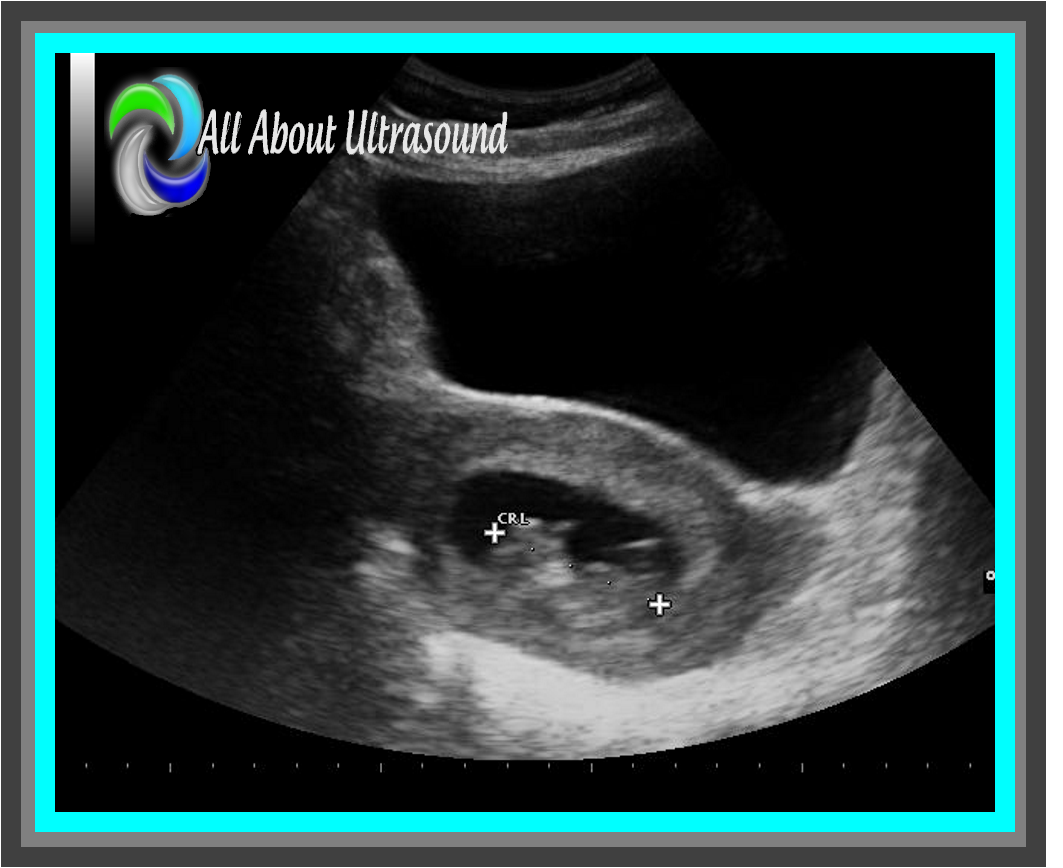
1. That Bladder! We have all seen the pregnant pee pee dance… While sometimes uncomfortable for our patients, this is the key to the first trimester transabdominal approach and also to 2nd trimester cervical measurements. Sometimes it just can’t be avoided, your patient is going to dance! However, in the 2nd trimester, it can be helpful for your patient to get what you need with the bladder full and to let them empty it out before completing the rest of your study. Often this can also improve the position of the fetus and help you to get better images with a relaxed mama and a relaxed baby!
Picture
Picture
2. Scanning and trying to find those ovaries can prove to be a challenge with a pregnant uterus! Even though it is not our focus, we can’t forget those ovaries! Making sure to image those along with the uterus and fetal images will ensure that you’re not missing potential pathology, but it can definitely be tough with that pregnant uterus in the way. Best methods are to bring the transducer laterally to the patient’s side and to scan in a transverse plane along the lateral side of the uterus, starting pretty high up and moving down toward the patient’s cervix. That transverse plane will give you an increased field of view and will allow you to identify structures more easily.

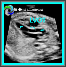
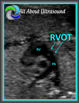
3. Outflow tracts… the sound of that might scare some sonographers who are not used to looking at the fetal heart or evaluating this on a regular basis, but it’s much easier than you might think. Remember there are two main blood vessels that exit the heart – the aorta and the pulmonary artery. That’s it… just two. So how do you tell which is which? Most of the time (unless in a case of transposition of the great arteries or double outlet right ventricle), the aorta exits the left ventricle and the pulmonary artery exits the right ventricle. It’s important to note that the aorta will arch around after it leaves the heart and extend into the thoracic/abdominal aorta. The pulmonary artery will typically split into two branches.

​Yes, there are many variations that can happen with cardiac pathology and unless you specialize in maternal fetal medicine or fetal echocardiography, you may not know the specifics of every congenital heart disease you might encounter, but if you know the basics and what that looks like, you should be able to know when it’s just not right and alert your reading physician. If you’re not scanning this on a regular basis, it would be a good idea to add this to your daily protocol.
Picture
Picture
Picture
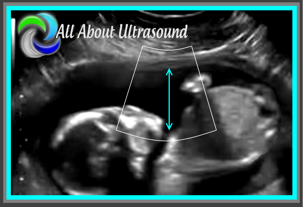
4. Amniotic Fluid Index – how do you know your measurements are accurate? Many sonographers are not aware of how to properly measure fluid for an AFI. One key tip is to scan with color Doppler flow on – this allows you to identify umbilical cord that might be floating within the pocket of fluid. Another best practice tip and proper way to perform AFI measurements is to be sure your patient is lying flat on the table and keep the probe/ultrasound beam perpendicular to the floor. Also, keep your measurements perpendicular as well. This helps to ensure that the fluid pocket is not falsely enlarged.
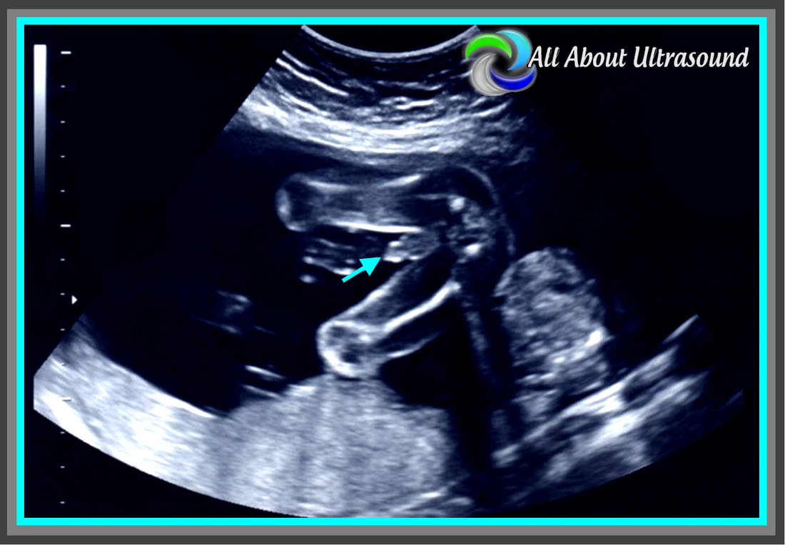
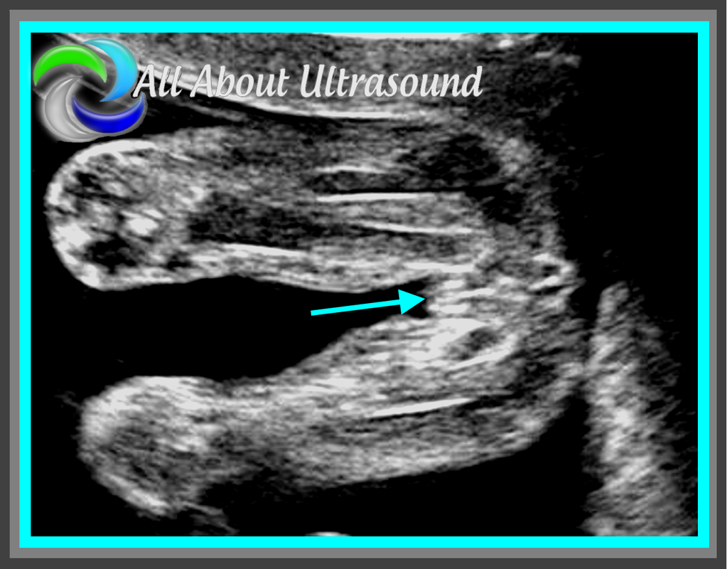
5. Gender! We’ve all been there, where it seems that everyone is there for the show. While somehow you ended up being the entertainment for the expectant parents, don’t forget how exciting it can be to know how to plan for your little one. Yes, it’s often a pain with the dad, grandma, grandpa, sister, brother, niece, nephew, the mailman, the neighbor, the sister’s boyfriend’s mother-in-law and everyone else trying to squeeze into the room to catch a glance of the precious cargo and whatever package the baby might be sporting… so take a deep breath, don’t lose it on grandma and remember to SMILE 😊. Oh and scanning, well you know how that goes…​ get those pesky diagnostic measurements and anatomy evaluations out of the way and  then... turtle or hamburger… take your pick!
Picture
Picture

​

Share

16 Comments

6/2/2017

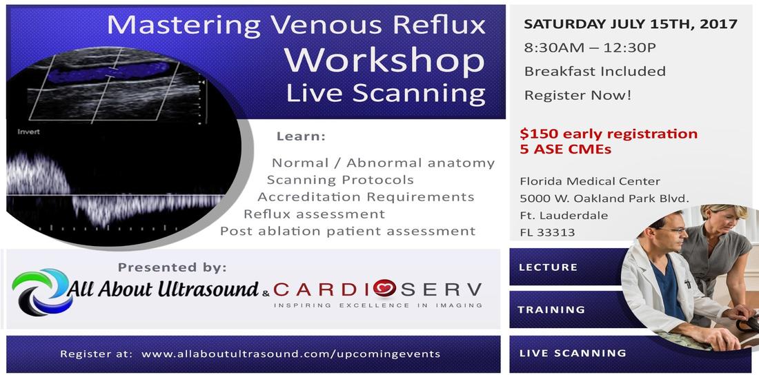
Mastering Venous Reflux - Live Scanning Workshop

0 Comments

Read Now
 
Picture
Don't miss our next LIVE SCANNING Event! In Sunny, Fort Lauderdale - July 15th 8:30am-12:30pm.

Together with the professionals from CardioServ, LLC we are hosting this amazing opportunity to learn venous insufficiency ultrasound. This course is for sonographers and physicians interested in learning correct techniques for performing and interpreting venous reflux.
  • Lecture and live scanning
  • Review normal and abnormal anatomy and pathology
  • Learn proper techniques and pitfalls to avoid
  • Diagnostic Criteria and intertpretation
  • Pre and Post ablation techniques
  • Instructors are credentialed experienced sonographers with real-life experience to teach and guide you
You will leave with tips to implement into your daily practice to improve the quality of your scanning and interpretation. Our goal is inspiring excellence in imaging. Let us inspire you to improve your venous reflux knowledge! Seating is Limited - Register Today!
Register Now

Share

0 Comments

5/28/2017

Ultrasound Investigators

0 Comments

Read Now
 
Unlike many other imaging modalities, scanning ultrasound is completely dependent on the sonographer, the angle of the probe and small little adjustments made to the machine settings. So often, it can be easy to just take that picture and move along. At All About Ultrasound, we want to make sure that sonographers like you have all the tools, tips, techniques and resources to not just pass a test or take a picture, but to excel at what you do. We want to give you the tools you need to transition from  being a picture taker to being a critical thinker and an investigator.  An image protocol is great for making sure that your images meet the basic standard criteria for accreditation and the basics of performing an exam, but it shouldn't end there. How do you investigate and give the physician what they need to make a diagnosis? How do you as the sonographer make a difference in the outcome of your patient? How do you learn and grow your skillset?

While we do offer many resources to add to your learning, we know that only those sonographers who truly seek out education and apply what they learn will ultimately be able to relate this back investigative patient care.  So, don't forget that there is a purpose for what you do. There is a reason you became a sonographer - the reason is your patient. Don't get so caught up in following the protocol that you don't ever go beyond it to investigate and piece the puzzle together to answer the clinical questions. You are not a picture taker - you are the eyes of the physician and without your investigative ultrasound skills, that doctor may not have what they need to treat the patient. So don't ever forget to seek out the answer, ask the questions, take that extra picture, take that extra Doppler sample and put that amazing sonographer brain to work. You are the eyes, the ears and the hands that can make a difference. It is so easy to get caught up in the day-to-day routine, but don't forget your WHY!

Share

0 Comments

4/19/2017

Gallbladder Wall Measurements

1 Comment

Read Now
 

Answer the age old question
Gallbladder measurements in Transverse or Longitudinal Plane?

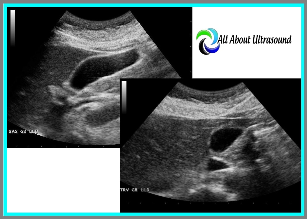
Depending on where you trained and who you trained with, you may have a different approach to gallbladder wall measurements than your colleagues. That age old question that has probably been around as long as sound waves... do you measure the gallbladder wall in a transverse or longitudinal plane? What is your protocol and why?
Picture

Share

1 Comment

4/4/2017

What's Your Diagnosis?

0 Comments

Read Now
 
What's your best diagnosis for this echocardiography clip?
​Note the jet seen in the parasternal short axis view and the apical long axis/3 chamber view.  
UPDATE:  4/5/2017

The video clip posted above shows a Coronary to Pulmonary Fistula. This type of communication can occur congenitally as well as post interventional and surgical cardiac procedures. According to a study sited by the Texas Heart Institute Journal, Coronary to Pulmonary communications, whether they are congenital or acquired, must be considered benign, in regard to their effect on coronary physiology.  Tex Heart Inst J. 2000; 27(4): 327–329.

See the video below with additional clips and images:

Share

0 Comments

2/8/2017

Ultrasound Registry Review

3 Comments

Read Now
 
Coming Soon! All About Ultrasound is making some BIG changes to our website. Get ready because over the next few weeks we will be adding full registry review courses to our lineup! Our Ultrasound Registry Review courses will be interactive with interesting and easy to learn training to prepare your for your ARDMS, CCI or ARRT ultrasound registry exam. No more boring ultrasound physics - Nope! Not here! Our review courses will help you learn what you need to know without the confusing lingo or difficult to follow outlines. Real talk for real people with real results. Learn ultrasound physics and more - Simple. Easy. Done! 
Picture

Share

3 Comments
<<Previous
Forward>>
Details
    Picture

    Making Waves​™

    All About Ultrasound presents Making Waves™, our ultrasound specific blog and newsletter. Join us here for ultrasound news, cases and more! 

    Suggestions or Comments?
    Tell us your opinion...
    [email protected]

    Don't FORGET YOUR MERCH!

    SHOP NOW
    Picture

    Archives

    May 2023
    April 2023
    November 2022
    July 2020
    June 2020
    January 2019
    December 2018
    February 2018
    January 2018
    July 2017
    June 2017
    May 2017
    April 2017
    February 2017
    January 2017
    September 2016
    August 2016
    July 2016
    June 2016
    May 2016
    May 2015
    April 2015
    March 2015
    January 2015
    July 2014
    July 2013
    March 2013
    February 2013
    November 2012
    April 2012
    April 2011
    March 2011
    January 2011
    November 2010

    Categories

    All
    Accreditation
    Case Studies
    Echocardiography
    General
    Job Search
    OB/GYN
    Physics
    Registries
    Salary
    Scanning Tips
    Ultrasound News
    Ultrasound Research
    Vascular
    Webinar

    RSS Feed


ALL RIGHTS RESERVED
​​©ALL ABOUT ULTRASOUND, INC. dba iHEARTECHO

TERMS OF SERVICE   ●   PRIVACY POLICY   ●   
SUPPORT
​
[email protected]

ARDMS® is a registered trademark of the American Registry for Diagnostic Medical Sonography and is not associated with this website.  
CCI® is a registered trademark of Cardiovascular Credentialing International and is not associated with this website.
​
​WEB DESIGN BY: SoundMedia
  • Home
  • Ultrasound Services
    • Ultrasound Accreditation
    • Competency Assessments
  • Ultrasound Education
    • E-Learning >
      • E-Learning & CME
      • Upper Extremity Duplex
      • Venous Insufficiency
      • Doppler Principles and Hemodynamics
      • Left Ventricular Diastology
      • Constrictive Pericarditis
      • Aortic Stenosis
    • Free Membership
    • On-Site Training
    • Scanning Quick Guides
  • Ultrasound Registry Review
    • Free Membership
    • Upcoming Events
    • Ultrasound Physics SPI
    • Advanced Cardiac Sonographer
    • Adult Echocardiography
    • Pediatric Echocardiography
    • Fetal Echocardiography
    • Vascular Ultrasound
    • Abdomen Ultrasound
    • OB-GYN Ultrasound
    • Breast Ultrasound
    • Group | Team Access
  • Ultrasound Jobs
  • Ultrasound Resources
    • Ultrasound Case Studies
    • Sonography Coloring Book
    • Ultrasound Worksheets
    • Ultrasound Protocols
    • Ultrasound Credentialing
    • ULTRASOUND SCHOOLS
    • Sonography News
  • Making Waves™ Blog
  • SONO SHOP
  • Newsletter
  • FAQ
  • About Us
  • Contact Us
  • FREE SIGN UP
  • LOGIN
  • iHeartEcho